摘要:
原发性视网膜色素变性(primary pigmentary degeneration of retina)亦称为色素性视网膜炎(retinitis pigmentosa, RP),是一组以进行性感光细胞及色素上皮功能丧失为共同表现的遗传性视网膜变性疾病,是一种比较常见的毯层-视网膜变性(tapetoretinal degeneration),以夜盲、进行性视野损害、眼底色素沉着和视网膜电图异常或无波为其主要临床特征,也是世界范围内常见的致盲性眼病。临床上“双眼视网膜色素变性”较为常见,“单眼视网膜色素变性”较为罕见,国内外杂志上也有报道。本篇病例报道介绍一例“典型的单眼视网膜色素变性”,从眼底照等临床上的检查结果以及基因检测分析此病例,提高“单眼视网膜色素变性”的诊断率,降低漏诊率。
Abstract:
Primary retinal pigment degeneration of retina, also known as retinitis pigmentosa (RP), is a group of hereditary retinal degenerative diseases with common expression of photoreceptor and pigment epithelium, a relatively common blanket layer - retinal degeneration (tapetoretinal degeneration) to night blindness, progressive visual field damage, fundus pigmentation and abnormal electroretinogram or no wave for its main clinical features, but also common worldwide blindness eye disease. Clinically, “binocular retinal pigment degeneration” is more common, “monocular retinal pigment degeneration” is relatively rare, and domestic and foreign magazines have also been reported. This case reports an example of “typical monocular retinal pigment degeneration”, from the fundus and other clinical findings and genetic testing analysis of this case, improving the "monocular retinal pigment degeneration" diagnosis rate, reducing the rate of missed diagnosis.
1. 病例报告
患者,男性,31岁,因“左眼逐渐视力下降5年余,伴夜盲”来我院就诊。患者5年前无明显诱因出现左眼视力下降,夜晚视物不清,因右眼视力正常,未引起重视,未到医院就诊。于1年前,左眼视力严重下降,遂到某医院进行就诊,2015.3.1检查视力:右眼1.0,左眼手动/眼前,予以“施图伦”眼药水点眼,“复方血栓通胶囊”口服三月后,视力未见好转。于2016.7.18来我院就诊。
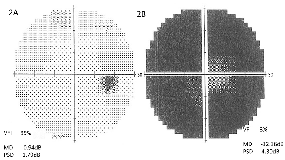
患者无炎症、外伤及手术史,无风湿性疾病,感染性疾病,药物中毒史,家族中无相似疾病。2016.7.18查视力:右眼1.0,左眼0.1,矫正不应;眼压:OD12.5 mmHg,OS14.6 mmHg。双眼角膜透明,前房清,深度可,瞳孔等大等圆,对光反射可,左眼扩瞳后可见晶体后囊下有锅巴样浑浊。右眼晶体透明。眼底照示左眼底窥视不清,视盘色蜡黄,界限清晰,视网膜动静脉血管变细,各个象限视网膜呈青灰色改变,周边视网膜及黄斑部有大量骨样细胞沉积(见图1)。左眼呈与眼底照相符的管状视野,右眼60˚视野无缺损(见图2)。光学相干断层扫描(OCT)示左眼视网膜视锥细胞及视杆细胞、视网膜色素上皮层萎缩性改变,右眼眼底照及OCT正常(见图3)。眼底荧光造影(FFA):左眼视网膜色素上皮萎缩呈斑状强荧光,广泛色素性荧光遮蔽,右眼荧光像无异常(见图4)。视网膜电流图(electroretinogran ERG)检查示左眼a b波幅值

Figure 1. Binocular fundus photography. 1A showed normal eye view of the right eye. 1B showed the left eye, visible optic disc hibiscus, retinal blood vessels thinner, a large number of peripheral retina-like cell deposition, and the entire retina was gray-gray change
图1. 双眼眼底合成照。1A显示右眼正常眼底照。1B为左眼眼底照,可见视盘蜡黄,视网膜血管变细,周边视网膜大量骨样细胞沉积,整个视网膜呈青灰色改变
平坦,右眼a b波幅值及峰潜时正常(见图5)。左眼视网膜诱发电位(VEP)示p100潜伏期延长(p100 138.5 ms),幅值降低(4.3 uv),右眼正常(参考值:9~45岁p100 95~115 ms,幅值5~25 uv) (见图6)。基因检测结果:突变位点为:GNAT1、CYP4V2、PDE6A、RBP3、USH1G、AHI1、CC2D2A,其中PDE6A、RBP3、

Figure 2. Binocular visual field. 2A showed normal visual field of the right eye. 2B showed the left eye visual field, visible visual field defect, a typical tubular field of vision
图2. 双眼视野检查。2A示右眼正常视野。2B示左眼视野,可见视野缺损,呈典型管状视野

Figure 3. Binocular optical coherence tomography (OCT). 3A showed normal OCT of the right eye. 3B showed the left eye OCT. The image showed the retinal cone cells and rod cells, retinal pigment epithelium atrophic changes
图3. 双眼光学相干断层扫描图像(OCT)。3A显示右眼正常OCT。3B为左眼OCT,图像显示视网膜视锥细胞及视杆细胞,视网膜色素上皮层萎缩性改变

Figure 4. Binocular fundus fluorescein angiography (FFA). 4A is normal eye FFA. 4B showed left eye retinal pigment degeneration of fluorescence angiography. Retinal pigment epithelial atrophy was patchy strong fluorescence, extensive pigmented fluorescence shielding
图4. 双眼眼底荧光造影图像(FFA)。4A为右眼正常FFA。4B为左眼视网膜色素变性的荧光造影,视网膜色素上皮萎缩呈斑状强荧光,广泛色素性荧光遮蔽

Figure 5. Eyes ERG check. 5A showed normal ERG of the right eye. 5B showed ERG examination of retinal pigment degeneration in the left eye. Dark adaptation fell
图5. 双眼ERG检查。5A示右眼正常ERG。5B示左眼视网膜色素变性的ERG检查。暗适应下降

Figure 6. Eyes VEP check. 6A showed normal VEP image of the right eye, p100 112.5 ms, amplitude 10.5 uv. 6B showed the left eye retinal pigment degeneration of the VEP image, p100 138.5 ms, amplitude 4.3 uv, p100 longer than normal, the amplitude decreased. (Reference value: 9 - 45 years old p100 95-115 ms, amplitude 5 - 25 uv)
图6. 双眼VEP检查。6A为右眼正常VEP图像,p100 112.5 ms,幅值10.5 uv。6B为左眼视网膜色素变性的VEP图像,p100 138.5 ms,幅值4.3 uv,p100较正常值延长,幅值降低(参考值:9~45岁p100 95-115 ms,幅值5~25 uv)。
USH1G位点突变与视网膜色素变性相关 [1] ,但由于是杂合突变,尚不能判定本病为其中一个基因突变所致。但临床症状比较典型,临床诊断为“左眼视网膜色素变性”。
2. 讨论
视网膜色素变性(retinitis pigmentosa RP)是视网膜感光细胞和色素上皮细胞变性所致的一组具有临床亚型的致盲性眼病,大部分于中年时期发病,视力急剧下降,伴发夜盲,是目前最常见的致盲性眼底病。西方国家RP发病率为0.025%~0.03%,国内发病率约0.024%~0.029%。RP的遗传方式包括常染色体显性遗传、常染色体隐性遗传,以及性染色体遗传,具有异质性。单眼视网膜色素变性临床罕见,文献报道其诊断标准 [2] :(1) 单眼有原发性RP的典型临床特征,视野及电生理检查呈现典型的RP特征。(2) 另一只眼完全正常,追踪5年以上仍未发病。(3) 排除炎症及外伤所致的继发性RP。本例病史虽然未追踪五年以上,但此患者从五年前既有视力下降,且所做的临床检查,包括眼底照,OCT,视野,FFA,电生理检查,高度怀疑其为“单眼视网膜色素变性”。为求进一步证实此诊断,我们会对病人做好随访工作。
原发性RP最早期的临床表现就是夜盲,并且呈进行性加重。RP的并发症包括白内障,经常会有晶体后囊下锅巴样浑浊表现 [3] 。近期也有报道RP会引发虹膜萎缩和青光眼 [4] 。典型的眼底改变包括视盘色蜡黄,视网膜血管变细,以及视网膜青灰色改变和骨样细胞沉积。另外临床上诊断RP还包括视野的检查,典型的RP病人视野向中心进行性缩小,晚期表现为典型的管状视野。RP病人眼底荧光素血管造影显示弥漫性的斑状强荧光,色素沉着处为荧光遮蔽。眼电生理检查,VEP示p100较正常潜伏期延长,振幅降低;Gauvin M [5] 等人对临床视网膜色素变性病人的长期研究获得了视网膜色素变性ERG的早期改变,RP病人ERG在早期即显示异常,表现为无波形,这为及早诊断RP提供了依据。
至今单眼RP的发病机制仍不明确,大多关于原发性RP发病机制的研究,双眼原发性RP病理机制首先是视杆细胞的损害,其后是视锥细胞亦收到损害 [6] 。感光细胞的损害主要是通过凋亡进行的,细胞神经营养因子缺乏也是凋亡的一个重要原因。基于单眼RP是单眼发病,大多数认为没有家族遗传病史,所以单眼RP的发病机制不能完全等同于原发性双眼RP。
本病目前尚无有效治疗方法,虽然有应用基因治疗的研究 [7] ,但效果并不明显,其他治疗方法还有药物治疗及视网膜移植 [8] ,但效果尚不理想 [9] 。
基金项目
国家自然科学基金资助项目(项目编号#81271015, #30872823, and #30801260)。