1. 引言
增殖性糖尿病视网膜病变(Proliferative diabetic retinopathy, PDR)是导致糖尿病患者视力丧失的重要原因。当出现严重的玻璃体积血(vitreous hemorrhage, VH)、牵拉性视网膜脱离(tractional retinal detachment, TRD)、视网膜前膜(epi-retinal membrane, ERM)时,须行平坦部玻璃体切除术(pars plana vitrectomy, PPV),清除玻璃体积血,剥除增殖膜,解除对视网膜的牵引并复位视网膜。但对于多数PDR患者,增殖性纤维血管膜与视网膜粘连紧密,术中剥膜极易发生医源性裂孔及大量出血,术中视野差、手术时间长、手术难度大,加之PPV术后并发症影响手术效果,最终患者视力往往得不到提高。研究表明,PDR患者眼内尤其是玻璃体内血管内皮生长因子(vascular endothelial growth factor, VEGF)含量大幅增高,其在PDR的发展恶化中起重要作用。抗VEGF治疗可有效阻止血管渗漏和新生血管的形成,减少PPV术中及术后并发症 [1] 。本文中我们将比较两组患者手术难易程度、手术并发症及其疗效。
2. 对象和方法
2.1. 对象
选取2014-12/2015-12我院收治的PDR患者23例26眼。其中单行PPV治疗的为对照组,康柏西普玻璃体腔内注射联合PPV的患者为试验组。患者纳入标准:(1) 经散瞳后眼底镜等检查确诊为增殖性糖尿病视网膜病变;(2) 视力明显下降;(3) 患者均签署PPV手术及康柏西普玻璃体腔内注射知情同意书;(4) 所有PPV手术及玻璃体腔注药均由同一手术医师完成。排除标准:(1) 存在其他眼部疾病;(2) 接受过激光治疗或存在其他眼部手术史;(3) 肝肾功能异常;(4) 存在心肌梗死、脑梗死、难以控制的高血压以及恶性肿瘤;(5) 妊娠或者哺乳期妇女。两组患者一般资料比较,差异无统计学意义(P > 0.05),具有可比性,见表1。
2.2.方法
2.2.1.手术方法
(1) 康柏西普玻璃体腔注射
术前抗生素滴眼液预防感染,所有操作均按照无菌操作要求完成,患者取仰卧位,表面麻醉,常规消毒铺无菌巾,于颞下象限睫状体平坦部进针,注射康柏西普0.5 mg/0.05ml于玻璃体腔内,棉签压迫5min,观察无渗漏,涂抗生素眼膏包眼。次日观察术眼前后节情况,测眼压。术后抗生素滴眼液点术眼 qid,直至行PPV治疗。
(2) 玻璃体切除术
手术前后抗生素滴眼预防感染,所有操作均按照无菌操作要求完成,患者取仰卧位,常规消毒铺无菌巾,利多卡因+罗哌卡因混合液球后阻滞麻醉,做23G显微切口切除玻璃体,根据术中情况行视网膜切开、复位、剥增殖膜、激光光凝、气液交换、硅油填充等。
2.2.2. 试验观察指标
记录两组患者手术前后 CFT、BCVA,手术时间,术中出血、医源性裂孔发生率,术后随访1年,记录4个月时CFT、BCVA及并发症的发生率。
2.2.3. 统计分析
采用SPSS 17.0统计学软件进行统计学分析。两组患者年龄、病程、术前空腹血糖、手术时间以χ̅±s表示,组间计量资料差异比较采用独立样本t检验,计数资料差异比较采用X2检验,以P<0. 05 为差异有统计学意义。
3. 结果
3.1. 两组患者手术情况的比较
本研究中试验组患者玻璃体腔内注射康柏西普后至行玻璃体切除术之前的时间内,我们观察到患者虹膜新生血管明显消退,玻璃体积血较前减轻,眼底新生血管膜也有不同程度消退。手术时间方面,试验组与对照组的平均手术时间相比:(75.64 ± 18.73) min VS (95.36 ± 14.74) min,差异有显著统计学

Table 1. Basic data of two groups
表1. 患者术前基本资料的比较
意义(t = −3.435, P = 0.001)。术中出现需要眼内电凝止血的严重、持续性出血眼数,试验组与对照组相比:2眼(14.3%) VS 7眼(58.3%),差异有统计学意义(X2 = 5.539, P = 0.019)。医源性裂孔发生率、硅油填充率、因复发玻璃体积血或视网膜脱离行2次手术率,试验组小于对照组,但差异并无统计学差异,填充硅油者均于术后3~6个月顺利取出硅油。随访期内因术眼白内障影响视力者均行白内障手术治疗。见表2。
3.2. 两组患者手术前后CFT变化的比较
本研究中试验组术后4 mo时黄斑中心凹厚度为(214.91 ± 30.15) μm,与术前黄斑中心凹厚度(441.92 ± 146.92) μm相比较,差异具有显著统计学意义(t = 5.019, P = 0.000)。对照组术后4 mo时黄斑中心凹厚度为(318.92 ± 156.17) μm,与术前黄斑中心凹厚度(452.23 ± 159.62) μm相比较,差异同样具有统计学意义(t = 2.152, P = 0.042)。两组患者术前黄斑中心凹厚度比较,差异无统计学意义,而试验组术后4 mo时黄斑中心凹厚度小于对照组,且差异具有统计学意义(P = 0.041)。见表3。试验组患者A随访期间OCT见图1。
3.3. 两组患者手术前后最佳矫正视力比较
本研究中视力采用ETDRS视力表测得,试验组术后4 mo时 BCVA 为(56.25 ± 22.90),与术前(20.65 ± 26.53)相比较,字母数增加,差异具有显著统计学意义(t = −4.543,P = 0.000)。对照组术后4 mo时BCVA为(38.57 ± 24.72),与术前(22.29 ± 21.78)相比较,字母数也有所增加。两组患者术前BCVA比较,差异无统计学意义(P = 0.851),而试验组术后4 mo时BVCA大于对照组,且差异具有统计学意义(P = 0.040)。见表4。
4. 讨论
PDR往往出现玻璃体出血、纤维增殖及牵拉所致的黄斑移位、黄斑水肿、视网膜脱离等。目前,PPV依然是PDR治疗的主流方法。该手术旨在切除积血的玻璃体,剥除新生血管膜,解除其对视网膜的牵拉,
Table 2. The operation of two groups
表2. 患者手术情况的比较
注:*为t值;#为X2值。
Table 3. The central foveal thickness of two groups before and after the operation
表3. 两组患者手术前后黄斑中心凹厚度变化的比较
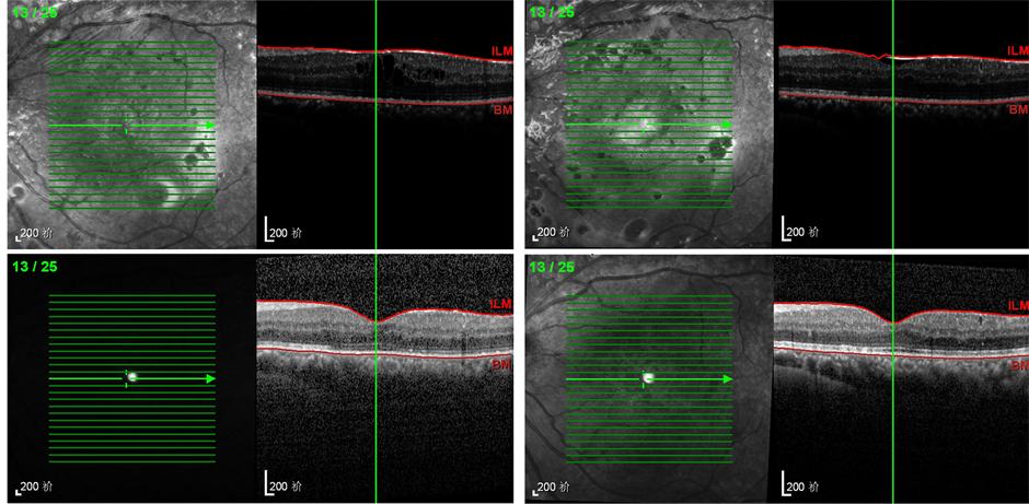 (左上:治疗前 右上:术后1mo 左下:术后3mo 右下:术后6 mo)
Figure 1. OCT of one patient in experimental group
图1. 试验组患者A随访期间OCT
Table 4. The comparison of the best corrected visual acuity in the two groups before and after the operation
表4. 两组患者手术前后最佳矫正视力的比较
最大程度上保留及提高患者的视功能 [2] 。但由于PDR患者的增殖膜与视网膜粘连紧密,术中剥膜困难,易出现医源性裂孔及反复、大量的出血。不仅手术难度大、耗时长,术后视力提高也不明显。因此,如何消退视网膜新生血管膜,成为PDR治疗的关键。近年来,我们发现VEGF在PDR的发生发展中,尤其在新生血管形成中起着重要作用。VEGF作为一种促有丝分裂因子和血管生成因子,能特异性刺激血管内皮细胞,导致其分裂、增生、游走 [3] ,并且通过增加血管的通透性、促进内皮细胞增殖 [4] 、增加内皮细胞对葡萄糖的转运 [5] 、改变细胞外基质、上调细胞间黏附分子1的基因表达等途径破坏血视网膜屏障,增加血管通透性,促使新生血管形成。因此,抗VEGF治疗受到越来越多的关注。在抗VEGF药物中,康柏西普是我国自主研发的一种重组融合蛋白,于2013年经我国CFDA 批准用于临床治疗。
本试验证实,抗VEGF治疗能为之后行PPV手术创造有力条件。术中我们发现:1) 试验组术中出血减少,这可能与康柏西普使新生血管膜收缩有关。2) 试验组新生血管膜更易剥除,这可能是由于康柏西普使血管通透性降低,改善视网膜充血,增强了视网膜的抗牵拉性,而且新生血管膜的收缩减少了与视网膜的粘附面积 [6] 。3) 术中出血减少,可使术中视野清晰、电凝止血的频率降低、手术时间减少,医源性裂孔的发生率也有所减少。术后因复发玻璃体积血或视网膜脱离再次手术率统计学差异并不显著,与先前学者的研究不符,可能与本研究样本量过小有关。
然而,抗VEGF治疗的作用是暂时而非持续性的 [7] 。有学者观察到抗VEGF治疗后,部分患者出现纤维组织增殖及TRD加重 [8] 。因此玻璃体腔内注射康柏西普后行PPV手术时间的选择显得尤为重要。已有研究发现,抗VEGF治疗后3天内,纤维血管增殖膜的抑制效果并不明显,10天后观察到的抑制效果确切,而多于14天手术效果差于14天前 [9] [10] 。本试验中,我们将手术时间定为玻璃体腔内注射康柏西普后6~8天,术中未发现有TRD加重的情况发生。
DME是导致糖尿病患者视力降低的主要原因之一。PPV可缓解部分黄斑区水肿,并提高大多数患者的视力 [11] [12] 。其机制可能包括:PPV可去除玻璃体-视网膜界面的牵引力,复位脱离的视网膜,解除玻璃体对黄斑的机械性牵拉;PPV可去除致病因子,改善视网膜的缺氧状态等。有动物实验证实,猴子眼球内持续高水平的VEGF可导致黄斑水肿的发生 [13] ,且患者眼内VEGF的表达水平与DME的严重程度呈正相关 [14] 。康柏西普作为一种VEGF受体,可抑制新生血管的形成,降低血管通透性,减少血管渗漏,从而减轻黄斑水肿,提高患者的视力。本试验证实:对照组与试验组术后CFT较术前均有所降低,BCVA也均较术前有所提高。且试验组的疗效不管从CFT还是BCVA都优于对照组。ETDRS标准对数视力表精确度高,重复性好,因此国际上,ETDRS视力测试已经取代snellen和sloan视力测试成为全球视力测试标准 [15] 。
综上所述,PDR围手术期玻璃体腔内注射康柏西普可有效地缩短手术时间,减少术中出血及术中术后并发症,增加手术的易行性,减少硅油填充率,同时可减轻黄斑水肿,使患者获得更好的最佳矫正视力,为临床治疗PDR开辟了一条新途径。但本试验样本数较少,随访时间较短,还需进一步增加样本量做更加深入地观察。