血氧水平依赖磁共振功能成像(functional MRI)以无创性、直观显示各分离脑区相互连接功能的特点 [1] 而被应用于人脑对相关事件反应的研究中。脑出血或外伤患者由于血肿的占位效应与神经损伤常引起各种神经功能障碍,而传统辅助检查只能定位病变位置和局部的脑功能,不能显示各个脑区之间的连接强度的变化。本研究将三位脑损伤后植物状态患者的脑fMRI图像,与三位正常同龄人作对比,显示患者脑部各功能区之间连接强度发生的变化,进一步探究影响患者觉醒与意识水平的脑区,为临床诊治提供参考。
1. 资料与方法
1.1. 实验对象
病例一:男,52岁,主诉“脑外伤后意识障碍3月”。患者于2013年5月21日车祸致脑外伤,立即出现意识丧失。送至浙江省温州市中医院CT提示右侧颞顶枕叶硬膜下血肿,中线结构移位。予行去骨瓣减压及血肿清除术,术后予积极治疗后病情稳定。2013年8月8日复查CT提示右侧硬膜下血肿清除术后改变,左侧丘脑、桥脑软化灶。既往有心动过速史。入院查体:BP 125/90 mmHg,P80次/分钟,植物状态。双侧瞳孔等大同圆,直径约3 mm,光反射灵敏,可见眼球水平运动。四肢肌肉轻度萎缩,四肢未见主动运动,双肩关节及髋关节活动受限,四肢肌张力略增高,双侧腱反射活跃,双侧Hoffmann征阳性,双侧掌颏反射阳性。诊断:脑外伤,植物状态,继发性癫痫。
病例二:男,48岁,主诉“脑外伤后意识障碍50天”。患者于2014年1月13日头部被人打击后昏迷,送至浙江省温州市中医院行头颅CT检查提示:左侧颅内出血,破入脑室,弥漫性脑水肿。当即给予“颅骨钻孔置管引流”。当日22点患者突发左侧瞳孔散大,予以“开颅血肿清除术 + 去骨瓣减压术”,术后入ICU治疗后病情平稳。既往有高血压病史。入院查体:BP 140/80 mmHg,P85次/分钟,植物状态,睡眠觉醒周期存在。四肢无自主运动,疼痛刺激肢体无屈曲反应,对光线声音刺激无明显反应。肩关节活动度受限,四肢腱反射亢进,双侧Hoffmann征阳性,Babinski征阳性,双侧掌颏反射阳性、吸吮反射阳性。诊断:脑外伤,弥漫性轴索损伤,植物状态,高血压病。
病例三:女,52岁,主诉“脑出血后意识障碍3月余”。患者于2014年11月5日中午突发头痛,后出现右手无力,言语不清,渐加重并有呕吐,送至浙江省温州市中医院行CT检查示“脑干出血”,行钻孔置管引流术,术后患者收入重症监护室,行气管切开。术后1月左右患者可睁眼,但对外界无反应。既往高血压病史。入院查体:植物状态,BP 130/100 mmHg,P68次/分钟。留置胃管,经口进水有呛咳。双侧瞳孔等大同圆,直径约3 mm,光反射存在。有不自主咀嚼样运动,下颌无偏移,角膜反射存在。双侧额纹、鼻唇沟对称。关节活动度无明显受限,仅左上肢偶可见自主活动,刺痛肢体可回缩。右侧Hoffmann征阳性,右侧Babinski征阳性,双侧掌颏反射阳性。诊断:脑干出血,植物状态,高血压病。
1.2. MRI扫描参数
脑损伤患者和正常对照的磁共振图像是在GE Discovery MR 750 3T磁共振扫描仪中采集的,采集过程中配合一个8通道的头部接收线圈。BOLD序列的参数为:TR/TE:2000/30 ms,翻转角:90˚,图像矩阵:64 × 64,层厚/层间距:4 mm/1 mm,FOV:230 × 230 mm2,共采集了31层。静息态fMRI扫描持续8分10秒,扫描过程中受试者保持睁眼,注视固定在头部线圈上的小镜子,镜子中有一个红色的十字注视点,受试者在扫描中尽量保持平静。本研究利用3D FSPGR序列得到受试者的高分辨率结构像。扫描参数为:TR/TE:7.8/3.0 ms,翻转角:20˚,反转时间:450 ms,FOV:240 × 240 mm2,层厚:2.0 mm,层间距:1.0 mm,分辨率:1 × 1 mm2。
1.3. 数据分析流程
本研究使用伦敦大学学院开发的统计参数图谱(Statistical Parametric Mapping 8, SPM8) [2] 进行BOLD数据的分析。BOLD数据的预处理主要包括以下步骤:
1) 头动校正:首先计算头动参数,包括三个旋转参数和三个平移参数。然后利用这些计算出来的参数进行头动校正,如果头动幅度较大,即平移超过2 mm,转动超过1˚,则该受试者的数据就会因为头动幅度过大影响数据质量而剔除。
2) 时间层校正:由于BOLD序列的k空间填充方式是蛇形填充,存在信号采集时间差,所以需要对功能像做时间层校正,以保证每层图像中神经元的活动时间一致。
3) 配准:将功能像与3D结构像做配准,并转换到MNI (Montreal Neurological Institute)空间上。
4) 归一化:将配准后的功能像与标准脑模板做归一化,以保证所有受试者的大脑图像都归一化到同一个标准脑上,便于后续的分析。
5) 空间平滑:用半峰全宽(Full Width at Half Maximum, FWHM)为4 mm的高斯内核对归一化后的功能像做截断为128秒的高通滤波。
本研究利用AAL模板将大脑分成90个脑区,并利用AAL模板得到了每个ROI的平均BOLD信号。本研究设置0.3为功能连接阈值,即连接系数大于0.3即认为有功能连接,连接系数小于0.3则认为没有功能连接。然后利用相关性分析得到90个脑区之间的功能连接矩阵。接下来,本文利用双样本双尾t检验对比了植物状态病人组和对照组之间脑区功能连接的差异。
2. 分析结果
三名植物状态病人的分析结果如图1,图2和图3所示。这是一个90*90的矩阵,每个小格子代表AAL模板中一个脑区与另一个脑区的连接强度。比如第一列第一个小格子代表左侧中央前回(PreCG.L)与自己的连接强度,强度为1。第一列第二个小格子代表左侧中央前回(PreCG.L)与右侧中央前回(PreCG.R)的连接强度。
三名正常对照组受试者的大脑连接矩阵如图4,图5和图6所示。
植物状态病人组和对照组的组分析结果如图7和图8所示。
双样本双尾t检验组分析结果如图9所示。
其中绿色色块代表二者无差异的连接,暖色色块(黄、浅红、深红)代表对照组强于病人组的连接,颜色越深,差异越大,冷色色块(青色、浅蓝、深蓝)代表病人组强于对照组的连接,颜色越深,差异越大。
这就是病人组和对照组大脑连接矩阵的差异,其中绿色色块代表二者无差异的连接,暖色色块(黄、浅红、深红)代表对照组强于病人组的连接,颜色越深,差异越大,冷色色块(青色、浅蓝、深蓝)代表病人组强于对照组的连接,颜色越深,差异越大。
3. 讨论
脑出血或脑外伤由于血肿压迫脑功能区、脑神经广泛损伤,常使患者进入昏迷状态,给家庭带来严重的经济与心理负担。早期有针对性的对特定脑区进行康复治疗,锻炼受损脑区相应功能,可更有效改善患者神经功能甚至恢复生活能力,帮助患者回归家庭与社会。
三位患者分别有左侧丘脑出血破入脑室、左侧颅内出血破入脑室、左侧脑干出血破入脑室的病史,
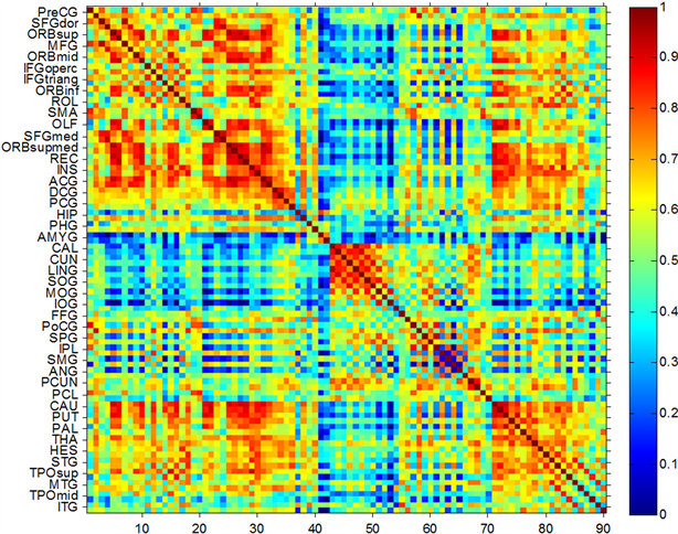
Figure 1. Brain connection matrix of the plant state of the subject one
图1. 植物状态受试者一的大脑连接矩阵

Figure 2. Brain connection matrix of the plant state of the subject two
图2. 植物状态受试者二的大脑连接矩阵
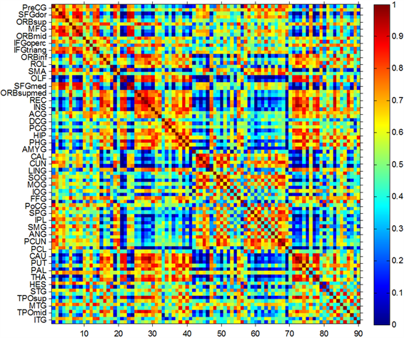
Figure 3.Brain connection matrix of the plant state of the subject three
图3. 植物状态受试者三的大脑连接矩阵
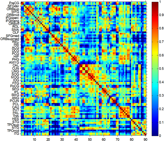
Figure 4. Brain connectivity matrix of subjects 1 in the control group
图4. 对照组受试者1的大脑连接矩阵
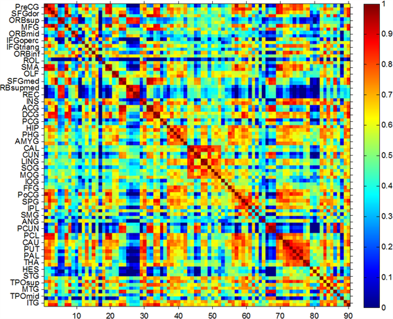
Figure 5. Brain connectivity matrix of subjects 2 in the control group
图5. 对照组受试者2的大脑连接矩阵

Figure 6. Brain connectivity matrix of subjects 3 in the control group
图6. 对照组受试者3的大脑连接矩阵
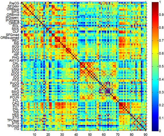
Figure 7. Analysis of 3 patients with plant status
图7. 3名植物状态患者组分析结果
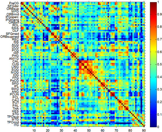
Figure 8. The results were analyzed in 3 normal subjects
图8. 3名正常受试者组分析结果
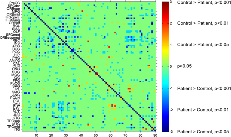
Figure 9. The difference in brain connectivity matrix between the patient and the control groups
图9. 病人组和对照组大脑连接矩阵的差异
恢复期生命体征平稳、对光反射正常、Horner征阴性,表现为明显的意识障碍、认知功能障碍、上下运动神经元瘫痪。本组脑出血患者出血病灶主要在左侧脑室附近,图像显示,除脑室周围基底神经节(尾状核、豆状壳核、豆状苍白球)、丘脑间的连接减弱外,其与额叶、扣带回间的连接也有减弱,而额叶(眶部额中回、脑盖部额下回、三角部额下回、眶部额下回、中央沟盖、辅助运动区、嗅皮质、眶内额上回)、扣带回附近(前扣带和旁扣带脑回、后扣带回、颞叶)、回直肌、脑岛相互之间的连接强度减弱更为密集与显著。左脑图像与右脑图像相比,损伤脑区有诸多重合,而损伤范围又小于右脑。研究者根据以上证据提出假设认为,三位患者意识障碍、认知障碍等临床表现与以上脑区相互连接减弱有极大关联(具有统计学意义),且以对侧(在此为右侧)大脑的损伤更为严重。据我们所知,国内还尚无探讨脑室出血后额叶、扣带回及附近脑区、基底神经节间相互连接功能强度减弱所致损害的文献报道。
4. 结论
基底节是埋藏在大脑皮质深部的灰质团块,是组成锥体外系的重要结构,过去认为,基底节与大脑皮质、丘脑、小脑等之间存在着纤维联系,在协同调节躯体随意运动、肌张力和姿势反射等方面起着重要作用,但近年来随着研究的深入,有越来越多的证据表明,基底节同时参与更多复杂行为的调节。基底节受损患者,常伴随语言功能下降、情绪认知障碍、记忆和注意障碍 [3] [4] [5] 等。额叶是大脑发育中最高级的部分,包括初级运动区、运动前区和前额叶区,与认知功能、注意力、记忆力等功能有着密切的关联,特别是前额叶区,关系最为密切。在感知觉整合障碍,处理问题能力、执行能力等方面起着重要的作用 [6] 。本研究病例提示,脑室出血所致意识障碍患者额叶间、额叶与基底神经节间的连接强度减弱,在原发病治疗的基础上,加强额叶与基底神经节相关功能的锻炼,对脑出血患者的康复治疗具有指导意义。