摘要:
报告一例经超声诊断的腹壁子宫内膜异位症患者。该患者有剖宫产手术史;手术切口瘢痕处结节,质硬,边界欠清,有触痛;有与月经相关的周期性切口瘢痕处疼痛等临床表现;超声表现:肿块位于腹壁切口处皮下软组织内,呈中低回声结节,边界欠清,形态欠规则,且彩色多普勒血流显像显示肿块内部及边缘可见血流信号,脉冲多普勒显示血流信号为低速高阻的动脉频谱。考虑符合腹壁子宫内膜异位症的诊断。术后病理诊断为子宫内膜异位症伴炎症反应。超声检查具有简便、快捷、经济、无痛苦、可重复性强等优点,并能动态观察病情变化,是诊断子宫内膜异位症的首选方法。
Abstract:
A patient with abdominal wall endometriosis diagnosed by ultrasonography was reported. The patient had a history of cesarean section. A hard and tender nodule with unclear border was found at the scar of surgical incision. There was periodic scar pain related with menstruation. Ultraso-nography showed the nodule located in the subcutaneous soft tissue of the surgical incision, with low echoes, unclear border and irregular shape. Color Doppler showed blood flow signals around and inside the mass, which were demonstrated as “low speed and high resistance” by Pulse Doppler. The data above supported the diagnosis of endometriosis. The pathologic diagnosis after surgery was endometriosis accompanied with inflammatory reaction. Ultrasonography had many advantages such as convenient, fast, inexpensive, painless, and repeatable. In addition, it could monitor the changes of diseases. Ultrasonography was the preferred method for diagnosis of endometriosis.
患者,女,34岁。主因腹壁切口瘢痕处疼痛3年,发现局部肿物1个月入院。患者3年前无明显诱因出现腹壁切口瘢痕部位顶端隐痛,呈间断性,尚可耐受,每于月经期疼痛加重,月经干净后疼痛缓解,未诊治。1个月前患者自行触及剖宫产瘢痕部位肿物,直径约3 cm,质硬,自感皮肤触痛,发现肿物增大。故就诊于我院,要求手术治疗。既往史:2003年剖宫产一足月女婴,2009年剖宫产一足月男婴。无高血压、冠心病及糖尿病史。否认肝炎、结核等传染病史。无外伤史,无输血史,无药物、食物过敏史。家族中无类似疾病及肿瘤、精神障碍等遗传性疾病史。
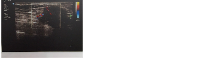
入院查体:T:36.6℃,Bp:125/79 mmHg。心肺四诊未见异常,腹平坦,下腹见长约8 cm的纵行剖宫产术后瘢痕,瘢痕部位顶端偏右侧、脐下可触及直径约3 cm的不规则、质硬肿物,边界尚清,表面光滑,有触痛,不活动。腹软,无压痛、反跳痛及肌紧张,叩鼓音,移动性浊音(−),肠鸣音正常存在。PV:已婚未产型外阴,阴道通畅,无充血及出血点,阴道分泌物量中,无异味,宫颈光滑,表面见那囊,子宫后位,正常大小,质中,活动可,无压痛,双侧附件区未见明显异常。辅助检查:腹壁体表包块彩超:腹前壁脐下紧邻皮下软组织层内可探及一中低回声结节,大小约21 × 27 × 20 mm,边界尚清,形态欠规则,内回声欠均,可见点状强回声。彩色多普勒及能量图:内部及边缘可见短棒状血流信号,可探及动脉频谱,PSV:22.6 cm/s,RI:0.77。超声印象:(见附录)腹壁中低回声实性结节(结合临床考虑子宫内膜异位症)。妇科腔内超声提示:腹部剖宫产切口上方皮下可见一大小约17 mm × 23 mm × 20 mm的低回声团块,边界欠清,未见明显血流信号。超声印象:腹部切口上方皮下低回声团块(考虑内异症)。根据:1) 患者有剖宫产手术史,2) 手术切口瘢痕处结节,质硬,边界欠清,有触痛,3) 有与月经相关的周期性切口瘢痕处疼痛等临床表现。结合超声表现:肿块位于腹壁切口处皮下软组织内,呈中低回声结节,边界欠清,形态欠规则;且彩色多普勒血流显像显示肿块内部及边缘可见血流信号,脉冲多普勒显示血流信号为低速高阻的动脉频谱。考虑符合腹壁子宫内膜异位症的诊断。完善相关检查后,查无手术禁忌,在腰硬联合麻醉下行腹壁子宫内膜异位症结节切除术。术后病理(BL198462):大体:结节一枚,直径3 cm,切面灰黄灰红,可见紫蓝色出血。病理诊断:(腹壁切口结节)子宫内膜异位,伴炎症反应。
讨论:具有活性的子宫内膜组织(腺体和间质)出现在子宫内膜以外部位时称为子宫内膜异位症(endometriosis, EMT),简称内异症 [1] 。子宫内膜异位症是妇科常见病、多发病,其好发部位为宫骶韧带、子宫直肠陷窝、卵巢以及盆腔腹膜的各个部位及盆腔脏器的表面,是一种良性肿瘤,但具有恶性肿瘤之远处转移和种植生长的特点。该病的发生机制至今尚未完全确定,目前学术界有种植学说、诱导学说等不同角度的学术解释,此外,遗传因素、免疫因素及炎症因素作为致病因素的观点也为临床医疗界所接受 [2] 。子宫内膜异位症发生部位大多在盆腔,但也可发生在腹部手术切口。子宫内膜异位到腹壁称为腹壁子宫内膜异位症(abdominal wall endometriosis, AWE) [3] 。近年来,由于社会因素的影响,剖宫产率逐年增加,术后发生的腹壁切口子宫内膜异位症亦随之增加,发生率为0.046% [4] 。腹壁切口内异症为种植学说提供了依据,有学者认为剖宫产术后,子宫内膜污染了手术切口,形成子宫内膜异位症。
子宫内膜异位症的基本病理变化为异位子宫内膜随卵巢激素变化而发生周期性出血,导致周围纤维组织增生和囊肿、粘连形成,在病变区出现紫褐色斑点或小泡,最终发展为大小不等的紫褐色实质性结节或包块。
本病临床表现:腹壁子宫内膜异位症常具有典型的临床表现,通常发生于妇产科手术后数月至数年出现周期性瘢痕处疼痛,在瘢痕深处扪及疼痛包块,症状与月经周期关系密切,经前疼痛加重,肿块增大,经后疼痛缓解,肿块缩小。由于肿块位于瘢痕组织内或与之关系密切,其活动度较差 [5] 。
腹壁切口子宫内膜异位症的超声声像图特征:1) 腹壁切口处探及不均质低回声肿块,形态欠规则、边界欠清晰或不清晰、无明显包膜回声。2) 肿块大小及回声随月经周期变化而变化,经前期及经期肿块增大,回声减弱;经后期肿块变小,回声略增强。3) CDFI (彩色多普勒)显示肿块内部血流信号稀少,周边部可见星点状或短线状血流信号,月经期肿块血流信号增多,血管增粗。4) PW (频谱多普勒)显示血流信号以动脉频谱为主,呈低速高阻型的特点 [6] 。
腹壁切口内异症诊断的依据:1) 既往手术史,包括剖宫产、经阴道分娩史及妇科手术史;2) 切口瘢痕周围出现肿块或结节肿块经前期和经期增大伴疼痛,经后缩小并疼痛缓解。多数患者据以上两点即可诊断。辅助超声检查可了解肿块大小、部位、性质、浸润深度和范围,有利于术前评估。CT显像上腹壁子宫内膜异位症可表现为实性或囊实性肿块,若病灶内有新鲜出血则可见片状或不规则形高密度影。由于病灶内含血供丰富的内膜腺体和乏血供的纤维组织,增强扫描可有不同程度的强化,但以轻度强化为主。侵及腹直肌筋膜层、肌层或腹膜时表现为肿块与相应结构粘连,局部筋膜或腹膜增厚 [7] 。
MRI信号表现比较复杂,增强扫描对于本病的诊断是必需的,增强扫描中病变明显强化,能清楚显示肿块对其向周围组织浸润的范围,增强扫描显示病变的范围明显大于平扫MRI,有利于在术前确定病变的大小及位置,增强扫描更能清晰显示其向周围组织浸润的深度,有利于手术前定位及彻底手术治疗 [8] 。近年来,CT、MRI等也用于术前检查,CT对软组织分辨率较低,且具有电离辐射,常常仅用于临床倾向不明确的患者。MRI具有软组织分辨率高,无电离辐射等优点,对于某些小病灶、尤其是含有血液成分的病灶具有很高的辨识能力。其符合率与超声相当,但由于其费用高,仅用于超声难以定位的患者。超声检查是诊断剖宫产术后腹壁切口瘢痕子宫内膜异位症的有效手段,再结合患者的剖宫产病史、临床表现,即可对内异症做出诊断,具有简便、快捷、经济、无痛苦、可重复性强等优点,并能动态观察病情变化,超声检查是目前诊断内异症的首选检查方法,为临床医生提供更多信息。

 附录
附录
期刊投稿者将享受如下服务:
1.投稿前咨询服务 (QQ、微信、邮箱皆可)
2.为您匹配最合适的期刊
3.24小时以内解答您的所有疑问
4.友好的在线投稿界面
5.专业的同行评审
6.知网检索
7.全网络覆盖式推广您的研究
投稿请点击:http://www.hanspub.org/Submission.aspx
期刊邮箱:acm@hanspub.org