感染性主动脉瘤的CT影像学分析与诊断
Computed Tomography Imaging Analysis and Diagnosis of Infective Native Aortic Aneurysm
DOI: 10.12677/acm.2024.1441289, PDF, HTML, XML, 下载: 32  浏览: 46 
作者: 霍正坤, 吴学君*:山东大学齐鲁医学院,山东 济南
关键词: 感染性主动脉瘤CT诊断动脉瘤列线图模型Infective Native Aortic Aneurysm CT Diagnosis Aneurysm Nomogram Model
摘要: 目的:通过感染性主动脉瘤和非感染性主动脉瘤的对照研究,寻找感染性动脉瘤特征性CT影像学表现,建立诊断模型。方法:纳入2013~2022年间于山东省立医院就诊的感染性动脉瘤病人87例,并应用倾向性评分匹配选择非感染性动脉瘤病例87例,对比两组间CT影像学表现,建立列线图影像学诊断模型。结果:囊性(52.9% vs. 16.1%),分叶状(40.2% vs. 3.4%)在感染性动脉瘤中更常见,管周积气(37.9% vs. 0%),积液(59.8% vs. 5.7%)和邻近组织感染(54.0% vs. 0%)在非感染性动脉瘤中罕见。感染性动脉瘤中的瘤壁钙化不能作为诊断依据(P = 0.158),多发动脉瘤在感染与非感染性动脉瘤组间无明显差异。依据囊性、分叶状、积气、积液、附壁血栓CT特征建立列线图诊断模型。结论:基于CT表现的列线图诊断模型具有较好的准确度和区分度。瘤体无钙化可能提示动脉瘤快速进展。多发性动脉瘤能否作为感染性动脉瘤提示特征还有待进一步讨论。
Abstract: Objective: To determine the CT imaging features of infective native aortic aneurysm (INAA) by comparing the infective native aortic aneurysm and non-infective aortic aneurysm (NIAA), and to build a nomogram diagnostic model. Methods: 87 cases of INAA patients during 2013~2022 were involved, and 87 cases of NIAA patients were selected by propensity-scoring match method. The difference of CT characteristics between two groups was compared. A nomogram diagnostic model based on the CT features was built. Results: Saccular (52.9% versus 16.1%), multilobular (40.2% versus 3.4%) aneurysm were more common in INAA. Periaortic gas (37.9% versus 0%), fluid (59.8% versus 5.7%) and adjacent infection (54.0% versus 0%) were the characteristic imaging features of INAA. There was no significant difference of multiple aneurysms between INAA and NIAA. A nomogram model based on features as saccular, multilobular, periaortic gas, fluid and mural thrombus was constructed. Conclusions: The nomogram model has a good sensitivity and specificity. Rapid progression can be indicated by the absence of calcification. The indication of the diagnosis of INAA by multiple aneurysms needs further discussion.
文章引用:霍正坤, 吴学君. 感染性主动脉瘤的CT影像学分析与诊断[J]. 临床医学进展, 2024, 14(4): 2247-2255. https://doi.org/10.12677/acm.2024.1441289

1. 引言

感染性原发主动脉瘤(Infective Native Aortic Aneurysm, INAA)临床进展较快,破裂率高,是一种罕见但致命的主动脉疾病。早期诊断对INAA降低死亡率、预防破裂至关重要 [1] 。该病临床表现不典型、血培养阳性率低、实验室检查特异性差,影像学检查常常是诊断INAA的有力证据 [2] [3] 。计算机断层扫描(CT)是临床实践中评估INAA的首选方式,其可以准确地显示INAA的位置及形态,明确破裂或先兆破裂,且通过定期复查可以发现是否存在动脉瘤快速进展(进展 > 5 mm/2周) [4] 。明确INAA的CT影像学特征,可以为INAA的临床诊断提供依据。

目前发表的关于INAA影像学的研究多为单组临床资料报告 [5] [6] [7] ,尚无纳入INAA与非感染性动脉瘤(Non-Infective Aortic Aneurysm, NIAA)的对照研究。本研究纳入87例感染性动脉瘤病例,并应用倾向性评分匹配选择NIAA患者作为对照,对比感染性动脉瘤与非感染性动脉瘤间的CT影像学差异,构建INAA的CT影像学诊断模型,并结合当前相关文献综述其相关的影像学研究进展。

2. 资料与方法

2.1. 资料收集

本研究纳入2013~2022年山东省立医院就诊的、出院诊断为“感染性主动脉瘤”的患者。排除标准包括 [8] :1) 非原发感染,如移植物感染、继发性主动脉瘘等;2) 虽符合诊断标准但可明确非感染性动脉瘤的。符合条件患者纳入观察组。非感染性动脉瘤患者应用倾向性评分匹配法(PSM),根据性别、年龄、动脉瘤位置(升主动脉及弓部、降主动脉、肾周腹主动脉、肾下腹主动脉或髂动脉)与观察组进行匹配,并纳入对照组。所有患者均签署知情同意书。

所有临床资料均自山东第一医科大学附属山东省立医院病历系统及检查结果系统提取,均为院内资料,包括:1) 基线资料(性别,年龄,基础疾病等);2) 临床表现;3) 实验室检查;4) CT影像学检查。

2.2. 统计分析

采用SPSS23.0统计学软件(美国IBM公司)进行分析。对照组应用倾向性评分匹配根据年龄、性别、动脉瘤位置三项评分1:1与观察组INAA患者匹配。连续性变量应用均值(标准差,范围)表示,二分类变量应用例数(百分比)表示。所有检验均为双侧检验,P值 < 0.05视为有意义,并计算95%可信区间。观察组及对照组组间差异应用Mann-Whitney U检验、χ2检验和独立样本t检验。采用单因素及多因素logistic回归分析筛选INAA的CT影像学危险因素,应用R 4.3.2绘制预测模型列线图,通过bootstrap自助抽样法做内部验证,绘制受试者工作特征(ROC)曲线、校准曲线(Calibration Curve)检验模型效能。

3. 结果

2013~2022期间,共1327例诊断为主动脉瘤的患者于山东第一医科大学附属山东省立医院血管外科就诊,其中87例被诊断为INAA并纳入观察组,87例倾向性匹配的非感染性动脉瘤(NIAA)患者被纳入对照组。INAA占全部动脉瘤患者的6.6%。

3.1. INAA的基线资料及临床表现

表1显示了87例INAA患者的基线资料及临床表现。患者平均年龄为67.1岁,其中77例(88.5%)

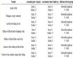
Table 1. Demographic data and clinical symptoms of 87 cases INAA patients

表1. 87例INAA患者基线资料及临床表现

注:免疫抑制或缺陷包括糖尿病、恶性肿瘤、肾衰竭、结核活动期、激素应用史、血液系统疾病、严重营养不良、风湿免疫疾病、梅毒、EB病毒感染;注2:其它系统感染包括肺炎、胆囊炎、泌尿系感染、皮肤感染、胃炎、阑尾炎、睾丸炎、肺脓肿、肝脓肿、胸膜炎、关节炎;注3:术前并发症包括下肢缺血、主动脉瘘、恶心呕吐、肠梗阻、腹泻、排尿困难、声带麻痹。

为男性。男性患者的平均年龄为66.3岁,女性患者平均年龄为73.7岁(P = 0.025)。其中合并高血压患者56例(64.4%),合并冠心病患者31例(35.6%),脑血管病患者15例(17.2%),呼吸系统疾病7例(8.0%)。51例患者合并免疫抑制或缺陷,其中糖尿病16例(18.4%),恶性肿瘤11例(12.6%),肾衰竭9例(10.3%)。存在疼痛的INAA病例约占77.0%,存在发热 > 38℃约占48.3%,其中同时存在发热及疼痛两大典型症状的患者仅有32例(36.8%)。存在破裂或先兆破裂的患者约占62.1%。

3.2. 对比INAA与NIAA的CT影像学表现

INAA组的平均直径为56.0 mm,显著小于NIAA组直径(60.7 mm, P = 0.035)。囊性或偏心性(46例,52.9% vs. 14例,16.1%,P < 0.001)及分叶状(35例,40.2% vs. 3例,3.4%,P < 0.001)是INAA中最常见的影像学特征,且在观察组中显著多于对照组(见表2)。33例INAA患者存在管周积气,52例患者合并积液,47例患者存在邻近部位感染,上述三项影像学特点在对照组中罕见(P < 0.001)。INAA组附壁血栓(45例,51.7% vs. 71例,81.6%,P < 0.001)和动脉瘤管壁钙化(36例,41.4% vs. 53例,60.9%,P = 0.01)均显著少于NIAA组。

INAA最常见于肾下腹主动脉段(50例,57.5%),其它还包括升主动脉段(4例,4.6%),主动脉弓(9例,10.3%),降主动脉段(18例,20.7%),肾周腹主动脉段(16例,18.4%),髂动脉段(24例,27.6%)。其中INAA组存在27例(31.0%)多发动脉瘤,与NIAA组(37例,42.5%)相比无显著差异(P = 0.12)。

Table 2. Comparison of laboratory tests and imaging features of INAA and NIAA

表2. INAA与NIAA组实验室检查及影像学特征对比

3.3. 建立列线图诊断模型

以是否诊断为INAA为因变量,以INAA与NIAA组间存在显著差异的CT影像学表现为自变量(瘤体直径、快速进展、囊性、分叶状、积气、积液、附壁血栓、钙化)进行二元logistic回归(表3)。将筛选出的独立危险因素(囊性、分叶状、积气、积液、附壁血栓)纳入列线图,构建CT影像学特征诊断INAA的列线图模型(图1)。

Table 3. Logistic regression of imaging features of INAA

表3. INAA影像学特征的logistic分析

Figure 1. Nomogram diagnostic model of INAA based on CT imaging features

图1. CT影像学特征诊断INAA的列线图模型

根据有无模型内的影像学特征,根据评分标准得到对应的分数。所有单项分数相加得到列线图总分,总分对应的预测概率为诊断为INAA的可能性。校准指数为0.91;ROC曲线下面积为0.91 (图2)。

4. 讨论

INAA临床表现不典型且缺乏特异性 [9] [10] 。因此影像学检查对于INAA的诊断至关重要,其中CT又是当前最常用的影像学检查方式。然而目前研究仅根据INAA的临床资料总结其影像学特征,尚无INAA与NIAA的对照研究。INAA的诊断则均结合临床表现、细菌培养结果与影像学表现。本研究通过进行INAA与NIAA的病例对照研究,寻找INAA的特征性CT影像学特征,构建基于影像学表现进行诊断的CT特征诊断模型。

INAA在CT中表现为囊性、偏心性、分叶状等特征的动脉瘤(图3),CTA可以精确描述INAA的瘤体形态(图4)。

Figure 2. The verification of nomogram model; (A) Area under curve was 0.91; (B) Calibration curve of nomogram, with C-index = 0.91

图2. 列线图模型的验证。(A) 曲线下面积(AUC)为0.91;(B) 列线图的校准曲线,校准指数(C-index)为0.91

Figure 3. Saccular aneurysm with periaortic exudation and ectopic gas (white arrow)

图3. 囊性动脉瘤合并管周渗出,管周积气(白色箭头)

本研究中,通过对比INAA和NIAA,证实快速进展、囊性、分叶状、积气积液、邻近感染等表现在INAA中显著多于NIAA。关于INAA管壁钙化的研究较少,一些研究报道瘤壁钙化在INAA中少见,也有一些研究报道多数INAA中存在钙化 [11] ,但均为单INAA组的临床资料报告。本研究对比INAA与NIAA的瘤壁钙化,发现INAA组内瘤壁钙化显著少于NIAA组(45例,51.7%)。动脉瘤的瘤壁钙化是在长期慢性炎症中形成的 [12] [13] ,其病程一般较INAA形成更长。因此我们推测这可能是由于感染导致瘤体进展较快,导致未形成钙化即成瘤。进一步logistic分析显示有无钙化不能作为INAA的诊断特征,故疑诊感染性动脉瘤患者的影像学表现无钙化不能作为诊断INAA的依据,但可能提示患者动脉瘤存在快速进展。

Figure 4. CTA of an INAA; (A) Saccular aneurysm close to the aortic bifurcation (white arrow); (B) Irregular multilobular aneurysm

图4. 感染性动脉瘤的CTA表现;(A) 近主动脉分叉处的左髂总动脉囊性动脉瘤(白色箭头);(B) 不规则分叶状动脉瘤

本研究中列线图模型校准曲线接近理想曲线,C-index = 0.91,AUC = 0.91,表示该模型准确度与区分度均较好。INAA的平均直径较NIAA小,但过半的INAA患者存在破裂或先兆破裂,提示即使小INAA也存在较高破裂风险。积气是INAA的特征性表现(图5),在列线图中评分约为100,即CT显示存在异位气体的患者,即使无其他特征,其动脉瘤感染的可能性也超过90% (图1)。囊性、分叶状动脉瘤是动脉瘤快速进展、病原体侵袭性的表现 [14] ,当存在这些表现时,即使患者未表现出INAA相关的临床表现或实验室检查,也应高度怀疑INAA的存在(>80%,图1)。多发动脉瘤在一些研究中被认为是INAA的特征 [11] [15] ,然而在本研究中,INAA组多发动脉瘤与NIAA无明显差异(P = 0.12),不能作为鉴别INAA与NIAA的依据。

Figure 5. Iliac INAA of same patient; (A) Preoperative ectopic gas (white arrow); (B) After EVAR intervention, a progression of ectopic gas (white arrow)

图5. 髂动脉感染性动脉瘤。(A) 患者术前可见管周少量积气(白色箭头);(B) 同一患者,腔内修复术后,积气增多(白色箭头)

主动脉瘘主要包括主动脉肠瘘、主动脉支气管瘘、主动脉食管瘘等,其影像学表现根据瘘管位置不同主要为造影剂分流或异位,可表现为下腔静脉在动脉期增强,或消化道、支气管等出现造影剂强化,主动脉瘘有时会出现对应症状,如黑便、咯血等 [16] [17] 。过半INAA累及肾下腹主动脉段(57.5%),但有文献报道主动脉瘘更好发于降主动脉段的INAA [18] 。

邻近感染中最常见的表现是管周组织感染渗出(图6),主要表现为静脉期造影剂边缘增强。腰大肌脓肿主要表现为水肿并单发或多发边缘强化脓肿。但是需要注意,破裂动脉瘤的CT影像可能会出现髂腰肌部位的血肿,与脓肿不易分辨。曾经有研究认为穿刺可能会导致感染复发或扩散 [19] [20] ,但近期的研究认为可以考虑穿刺以明确性质 [8] 。椎体感染可以是椎骨或椎间盘破坏,PET/CT也可以在早期未有器质性破坏时即提示椎体感染。

Figure 6. Periaortic exudation of INAA; (A) Iliac INAA with exudation; (B) Postoperative exudation around the stent of an abdominal INAA

图6. INAA管周组织渗出。(A) 髂动脉感染性动脉瘤合并管周渗出;(B) 腹主动脉感染性动脉瘤支架术后管周积液

5. 总结

本研究首次通过病例对照研究对比INAA与NIAA的影像学表现。基于囊性、分叶状、积气、积液、附壁血栓的CT影像学特征,本研究亦首次构建了INAA的影像学特征列线图诊断模型,模型准确度与区分度均较好,可以依据该模型及时诊断INAA并早期处理。本研究讨论了瘤壁钙化在INAA中的提示意义,无钙化的INAA可能提示动脉瘤快速进展。多发动脉瘤能否作为INAA的诊断依据仍然有待进一步讨论。本研究亦存在局限性。样本量在INAA的临床研究中较高,但受限于其低发病率仍然不足,影响结论的统计学意义。同时研究对象均来自单中心,可能对其结论的推广产生影响。

NOTES

*通讯作者。

参考文献

[1] Ballaith, A., Raffort, J., Rajhi, K., et al. (2022) Mycotic Aortic and Left Iliac Ruptured Aneurysm Due to Escherichia Coli: A Case Report and Literature Overview. Acta Chirurgica Belgica, 122, 56-62.
https://doi.org/10.1080/00015458.2020.1753146
[2] Plotkin, A., Magee, G.A., Elsayed, R.S., et al. (2020) Methicillin-Resistant Staphylococcus aureus Portends a Poor Prognosis after Endovascular Repair of Mycotic Aortic Aneurysms and Aortic Graft Infections. Journal of Vascular Surgery, 72, 276-285.
https://doi.org/10.1016/j.jvs.2019.08.274
[3] Sorelius, K. and Di Summa, P.G. (2018) On the Diagnosis of Mycotic Aortic Aneurysms. Clinical Medicine Insights: Cardiology, 12.
https://doi.org/10.1177/1179546818759678
[4] Wanhainen, A., Verzini, F., Van Herzeele, I., et al. (2019) Editor’s Choice—European Society for Vascular Surgery (ESVS) 2019 Clinical Practice Guidelines on the Management of Abdominal Aortoiliac Artery Aneurysms. European Journal of Vascular and Endovascular Surgery, 57, 8-93.
https://doi.org/10.1016/j.ejvs.2018.09.020
[5] Sorelius, K., Wanhainen, A., Furebring, M., et al. (2016) Nationwide Study of the Treatment of Mycotic Abdominal Aortic Aneurysms Comparing Open and Endovascular Repair. Circulation, 134, 1822-1832.
https://doi.org/10.1161/CIRCULATIONAHA.116.024021
[6] Dang, Q., Statius, V.E.R., Wever, J.J., et al. (2020) Nationwide Study of the Treatment of Mycotic Abdominal Aortic Aneurysms Comparing Open and Endovascular Repair in the Netherlands. Journal of Vascular Surgery, 72, 531-540.
https://doi.org/10.1016/j.jvs.2019.09.060
[7] Luo, Y., Zhu, J., Dai, X., et al. (2018) Endovascular Treatment of Primary Mycotic Aortic Aneurysms: A 7-Year Single-Center Experience. Journal of International Medical Research, 46, 3903-3909.
https://doi.org/10.1177/0300060518781651
[8] Sorelius, K., Wyss, T.R., Adam, D., et al. (2023) Editor’s Choice—Infective Native Aortic Aneurysms: A Delphi Consensus Document on Terminology, Definition, Classification, Diagnosis, and Reporting Standards. European Journal of Vascular and Endovascular Surgery, 65, 323-329.
https://doi.org/10.1016/j.ejvs.2022.11.024
[9] Shirasu, T., Kuno, T., Yasuhara, J., et al. (2022) Meta-Analysis Finds Recurrent Infection Is More Common after Endovascular than after Open Repair of Infected Abdominal Aortic Aneurysm. Journal of Vascular Surgery, 75, 348-355.
https://doi.org/10.1016/j.jvs.2021.07.240
[10] Sorelius, K., Budtz-Lilly, J., Mani, K., et al. (2019) Systematic Review of the Management of Mycotic Aortic Aneurysms. European Journal of Vascular and Endovascular Surgery, 58, 426-435.
https://doi.org/10.1016/j.ejvs.2019.05.004
[11] Jutidamrongphan, W., Kritpracha, B., Sorelius, K., et al. (2022) Features of Infective Native Aortic Aneurysms on Computed Tomography. Insights Imaging, 13, Article No. 2.
https://doi.org/10.1186/s13244-021-01135-x
[12] Joshi, N.V., Elkhawad, M., Forsythe, R.O., et al. (2020) Greater Aortic Inflammation and Calcification in Abdominal Aortic Aneurysmal Disease than Atherosclerosis: A Prospective Matched Cohort Study. Open Heart, 7, E1141.
https://doi.org/10.1136/openhrt-2019-001141
[13] New, S.E.P. and Aikawa, E. (2011) Cardiovascular Calcification—An Inflammatory Disease. Circulation Journal, 75, 1305-1313.
https://doi.org/10.1253/circj.CJ-11-0395
[14] Zhang, N., Xiong, W., Li, Y., et al. (2021) Imaging Features of Mycotic Aortic Aneurysms. Quantitative Imaging in Medicine and Surgery, 11, 2861-2878.
https://doi.org/10.21037/qims-20-941
[15] Sorelius, K., Mani, K., Bjorck, M., et al. (2014) Endovascular Treatment of Mycotic Aortic Aneurysms: A European Multicenter Study. Circulation, 130, 2136-2142.
https://doi.org/10.1161/CIRCULATIONAHA.114.009481
[16] Lakhani, D.A., Sharma, S.A., Kutayni, H., et al. (2021) Primary Aortoenteric Fistula: A Case Report and Brief Review of the Literature. Radiology Case Reports, 16, 3549-3553.
https://doi.org/10.1016/j.radcr.2021.08.044
[17] Tournaye, E., Hollering, P., De Roover, D., et al. (2022) Staphylococcus aureus Sepsis and Hemoptysis as Messengers of a Rather Impractically Located Mycotic Aneurysm. Acta Chirurgica Belgica, 123, 430-435.
https://doi.org/10.1080/00015458.2022.2030127
[18] Bento, R., Rodrigues, G., Alves, G., et al. (2023) Mycotic Aortic Aneurysm: A Ticking Time-Bomb. Angiologia e Cirurgia Vascular (Sociedade Portuguesa de Angiologia e Cirurgia Vascular), 19, 47-49.
[19] Jonker, F.H., Heijmen, R., Trimarchi, S., et al. (2009) Acute Management of Aortobronchial and Aortoesophageal Fistulas Using Thoracic Endovascular Aortic Repair. Journal of Vascular Surgery, 50, 999-1004.
https://doi.org/10.1016/j.jvs.2009.04.043
[20] Roldan, C.J. (2012) Penetrating Atherosclerotic Ulcerative Disease of the Aorta: Do Emergency Physicians Need to Worry? Journal of Emergency Medicine, 43, 196-203.
https://doi.org/10.1016/j.jemermed.2011.01.026