1. 引言
犬睾丸恶性精原细胞瘤(Malignant Seminoma)是最常见的睾丸肿瘤,常发生于没有下降的睾丸。肿瘤细胞来源于精原细胞。国内外关于犬睾丸恶性精原细胞瘤的报道较少[1] 。现将本实验室接诊的一例犬睾丸弥散型恶性精原细胞瘤病例报告如下。
2. 临床资料
患病动物为8岁雄性吉娃娃犬,右侧腹股沟处曾发生隐睾,一个月前发现睾丸迅速变大,变硬,由动物主人送诊就医。兽医经去势手术治疗后将睾丸切除后固定送检至北京观赏动物医院。切除后的睾丸表面有一层包膜,质地较硬,表面凹凸不平,呈白色椭圆形,直径约为2~3 cm。
3. 检测方法
3.1. 送检材料
10%福尔马林固定液固定的睾丸组织,所有动物实验均按照北京市实验室动物管理办公室批准的北京市福利实验动物和伦理审查指导方针进行实验。
3.2. 制作切片
将组织按照酒精脱水,二甲苯透明,石蜡浸蜡和包埋,切片,贴片和烘片后制作成厚度为5μm的切片,对切片进行常规的H&E 染色后进行光镜观察。
3.3. 图像获取
OLYMPAS U-S R G数码显微镜观察并分别在低倍(100×)和高倍(400×)视野下采图。
4. 组织病理学检查结果
低倍镜观察:增生的细胞实质由大小不等的小叶状结构组成,睾丸小叶内含形态单一的细胞群弥漫性增生,成带状、块状和条索状排列,睾丸小叶之间有数量不等的结缔组织所分割,可见部分血管充血(图1(a))。
高倍镜观察:可见增生的细胞体积较大但大小形态较为一致,细胞中央为大的圆形细胞核,核膜清晰,核仁明显,细胞核被细条状染色质贯穿,可见核分裂像。胞浆丰富并且呈透明状(图1(b)),支持性间质内含数量不等的类淋巴细胞(图1(c))。
诊断结果为:睾丸弥散型恶性精原细胞瘤。治疗结果:经去势手术治疗后预后良好。
5. 讨论
犬的精原细胞瘤主要表现为睾丸的增生,肿胀。老龄犬较常见。发生在单侧或双侧睾丸,右侧睾丸发病率略高于左侧。常发生于没有下降的睾丸,易发犬种包括拳师犬,吉娃娃犬,波美拉尼亚犬,贵妇犬等。肿瘤大小不等,呈灰白色,质地较硬,但硬度小于睾丸支持细胞瘤[2] ,表面凹凸不平。肿瘤的部分区域也会发生出血和坏死[3] 。根据肿瘤的组织学特征可将其分为小管内型和弥散型。小管内型的肿瘤细胞常发生在早期,增多的肿瘤细胞填充在输精管内,替代正常的精原细胞和支持细胞。这种肿瘤细胞体积较大,呈多角型,边缘较锐。细胞核清晰可见,核仁明显呈棒状,有丝分裂像多见且数目不等,胞质透明,有明显的胞膜。伴有淋巴细胞浸润,也可形成淋巴滤泡。偶尔可见浆细胞和嗜酸细胞。肉芽肿性反应和纤维化常见,有时甚至不易辨认肿瘤。弥散型的肿瘤细胞不仅局限于输精管内,常呈片状分布。分布于新生肿瘤细胞间的坏死的肿瘤细胞呈现“星空样”变。90%的精原细胞瘤为典型或经典型,其余由罕见类型组成。如精母细胞性精原细胞瘤(Spermatocytic Seminoma),间变性精原细胞瘤(Anaplastic Seminoma)和伴有合体细胞滋养层巨细胞的精原细胞瘤[4] 。精原细胞瘤常采用手术治疗,摘除睾丸后,
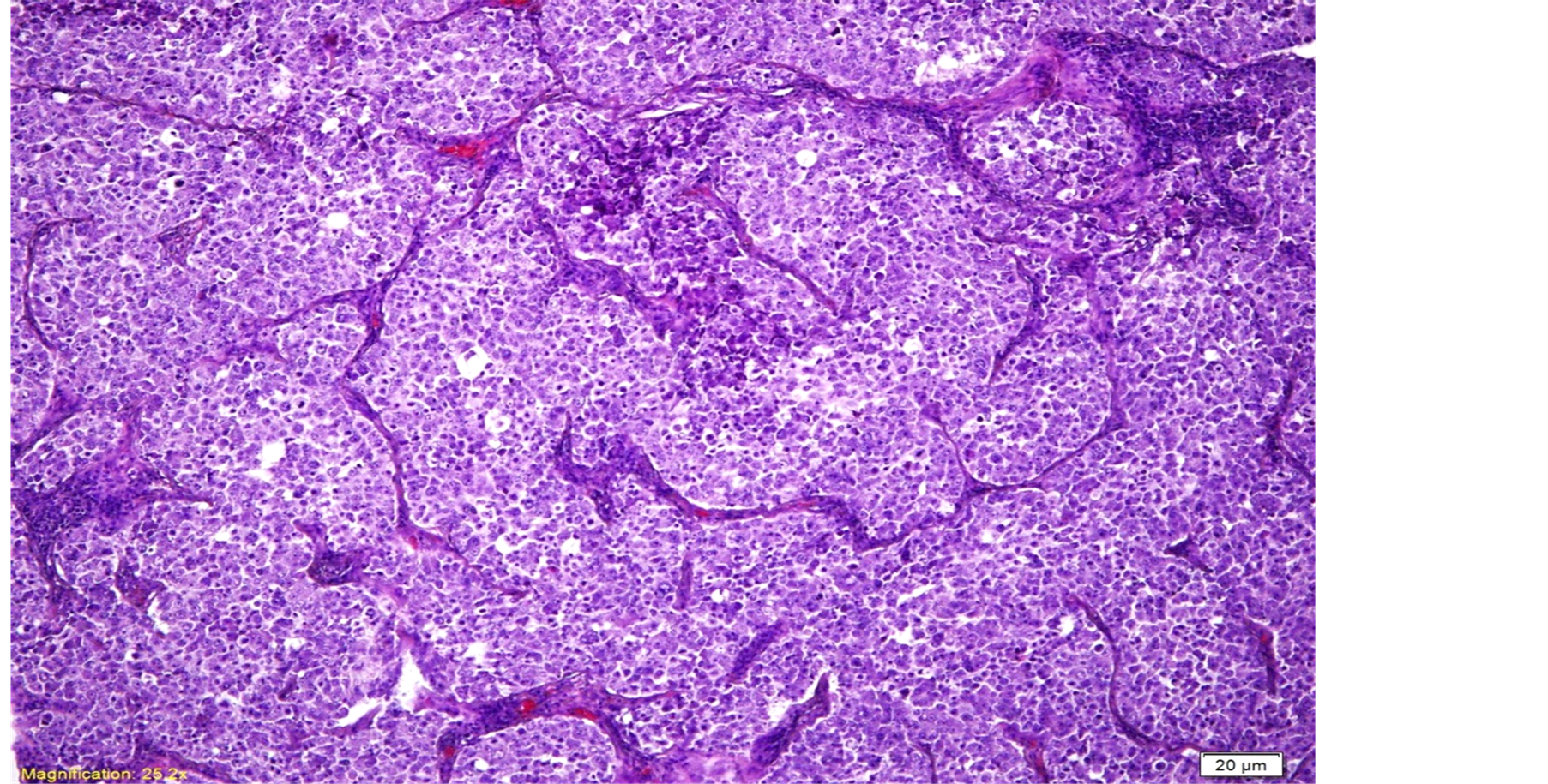
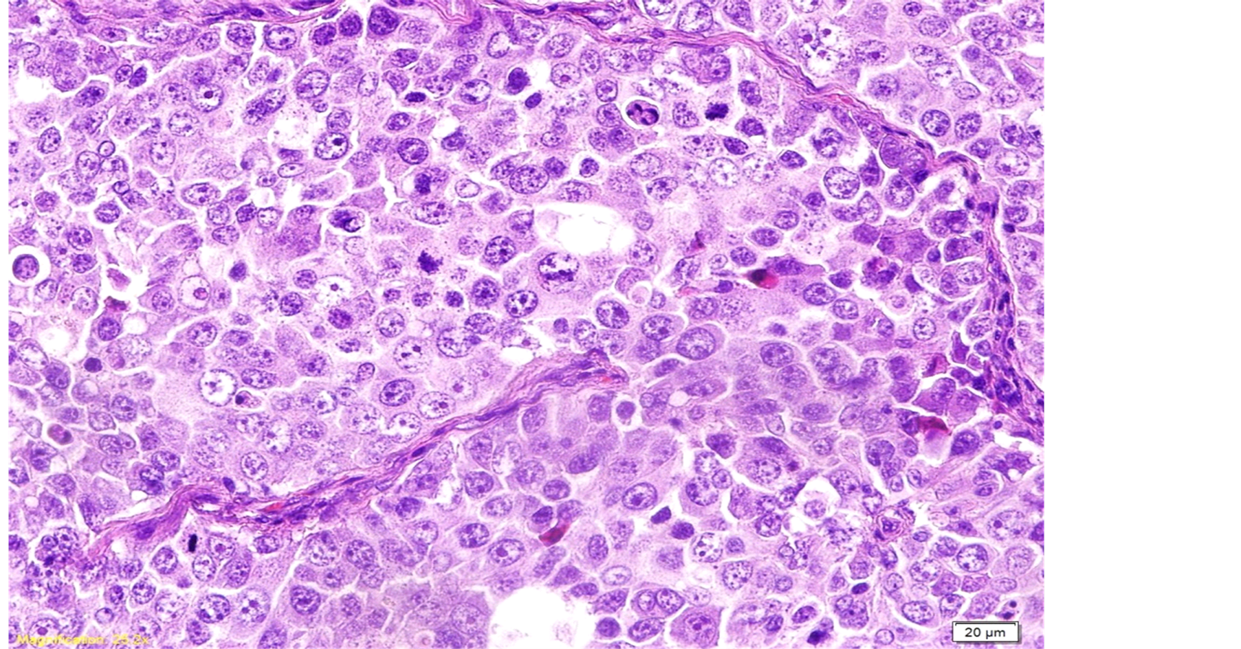
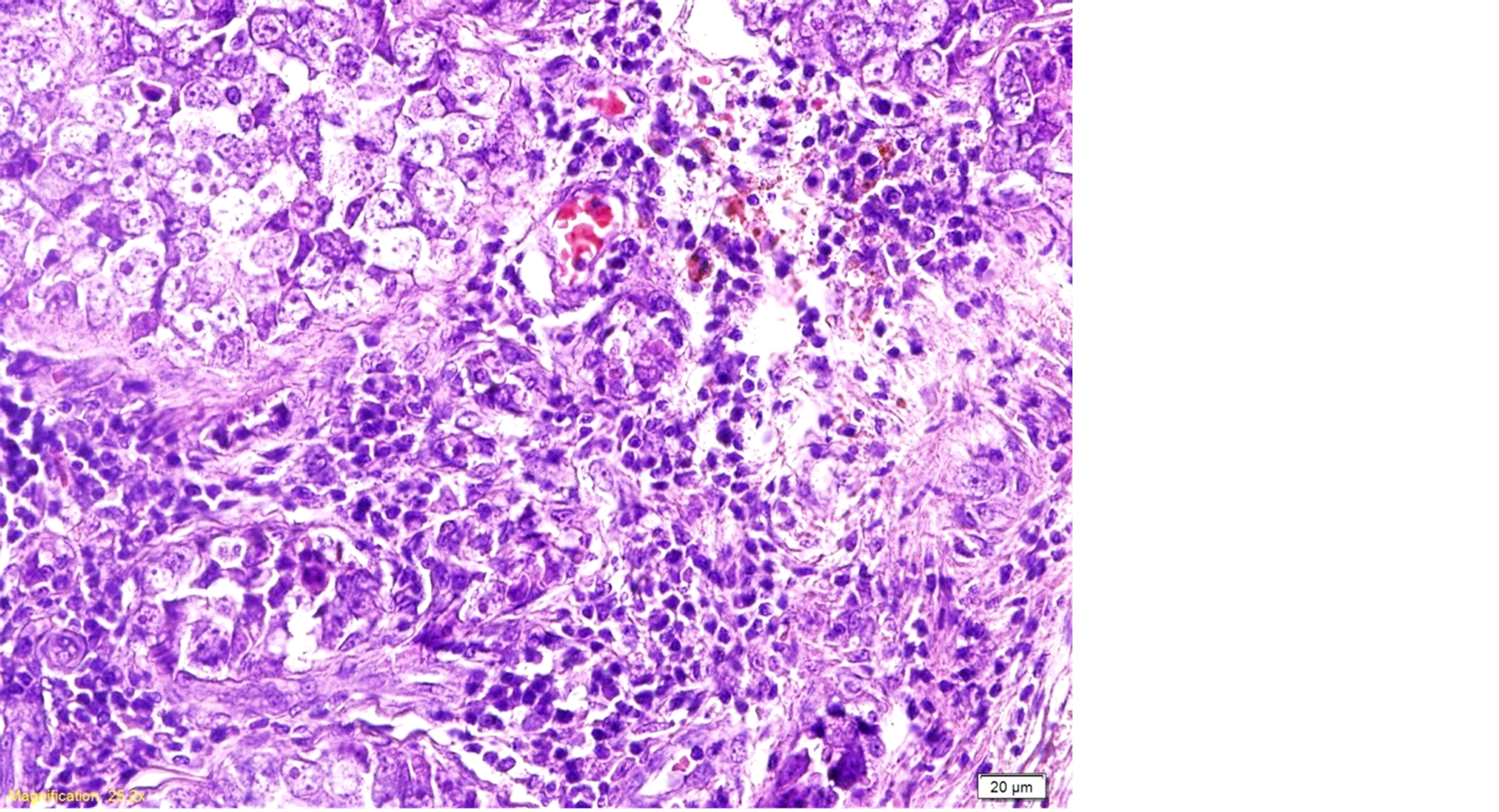
(a) 睾丸小叶内含形态单一的细胞群弥漫性增生,呈带状、块状和条索状排列(100×, H & E);(b) 增生的细胞形态一致,胞核深染,核仁明显,可见核分裂像;胞浆丰富并且呈透明状(400×, H & E)(c) 支持性间质内含数量不等的类淋巴细胞,部分血管充血、出血(400×, H & E)
Figure 1. Histopathology images
图1. 组织病理学图像
预后良好。
精原细胞瘤需和胚胎性癌(Embryonal Carcinoma)以及淋巴瘤(淋巴瘤)相区分。相对于精原细胞瘤来说细胞和核的多形性更为明显、分裂像更活跃。有些区域出现乳头状,假腺状结构以及合体排列细胞。淋巴瘤通常是大细胞型,其特点为肿瘤细胞在曲细精管间的间质内浸润生长。瘤内没有恶性生殖细胞增生,而且常无纤维化肉芽肿性反应。淋巴瘤比精原细胞瘤更常累及白膜,附睾和精索[5] 。
基金项目
国家科技支撑计划,十二五,课题编号:2011BA115B01。