摘要:
目的:眼底自发荧光(fundus autofluorescence, FAF)技术是近年来应用到临床的一项视网膜影像学技术,其可用于快速、无创地诊断和监测包括后葡萄膜炎在内的很多眼底疾病。本文将探讨FAF在后葡萄膜炎的影像特征及其临床应用价值。方法回顾性分析9例(15只眼)后葡萄膜炎患者的临床资料,分别行FAF、光学相干断层扫描(optical coherence tomography, OCT)、荧光素钠眼底血管造影(fundus fluorescence angiography, FFA)或吲哚青绿脉络膜血管造影(indocyanine green angiography, ICGA)等检查,对比FAF与眼底血管造影(FFA或ICGA)在监测黄斑水肿中的效果分析;对比后葡萄膜炎治疗前后眼底自发荧光的表现并进行评价分析。结果:1) 9例(15眼)后葡萄膜炎患者行FAF检查显示12只眼黄斑区呈范围大小不一的异常强荧光,而FFA/ICGA检查显示其中9眼黄斑区呈强荧光积存,同时OCT检查显示其中10眼黄斑区神经上皮层均有不同程度隆起增厚。2) 9例(15眼)后葡萄膜炎患者在治疗前后患眼视力有不同程度提高或保持不变(11眼视力不同程度提高,4眼视力保持不变),其相应FAF检查显示异常高荧光减弱,范围稍有缩小,同时异常低荧光区域不变或稍有扩大。结论:FAF在评估后葡萄膜炎黄斑水肿及评价后葡萄膜炎治疗前后疗效分析等方面具有简单无创、临床应用性强且随访性强的特殊应用价值。
Abstract:
Objective: Fundus autofluorescence is a new developed technique in opthalmoscopic imaging in recent years. It can monitor and diagnose various kinds of fundus diseases including posterior uveitis quickly and noninvasively. In this paper, we discussed the image feature and clinical ap-plication about fundus autofluorescence in posterior uveitis. Methods: We retrospectively analyzed 9 patients (15 eyes) suffering from posterior uveitis, and some eximanations were given to all of the patients, such as fundus autofluorescence (FAF), optical coherence tomography (OCT), fundus fluorescence angiography (FFA) or indocyanine green angiography (ICGA). First, we compared FAF with FFA or ICGA in evaluating macular edema; then we observed and analysed treatment effect by comparing manifestations before and after the treatment for posterior uveitis in FAF. Results: 1) 12 eyes of 9 patients (15 eyes) suffering from posterior uveitis show abnormal high fluorescence with irregular size in FAF. However, 9 eyes of them show high fluorescence in FFA or ICGA. Meanwhile, 10 eyes show that their retinal neurosensory layer in fovea thickens obviously in OCT. 2) The eyesight of 9 patients (15 eyes) suffering from posterior uveitis improves varying degrees or remains unchanged (the eyesight of 11 eyes improves and 4 of them remain unchanged), and their corresponding FAF expression shows that the intensity and extent of abnormal high fluorescence both reduce; the extent of low fluorescence remains unchanged or extends a little. Conclusions: There are special applications for FAF to evaluate macular edema and estimate treatment effect for posterior uveitis. Other than greater compliance with follow-up, it’s also non-invasive and easy to operate.
1. 引言
后葡萄膜炎[1] 是累及脉络膜、视网膜、视网膜血管及玻璃体的炎症性疾病,主要有脉络膜炎、视网膜炎、脉络膜视网膜炎、视网膜脉络膜炎、视网膜血管炎等。常见的临床表现有:玻璃体炎症细胞、混浊;局灶性浸润病灶;血管炎(鞘、闭塞、出血);黄斑水肿;渗出性网脱;增殖性视网膜病变;玻璃体积血等。以往对后葡萄膜炎的诊断及监测主要依赖于FFA或ICGA及OCT等,而FAF[2] [3] 作为一种非侵入、不接触、无损伤、快速并可重复地观察及监测包括后葡萄膜炎在内的很多眼底疾病的新应用,已经开始发挥它独特的作用,并越来越多地受到眼科医生及医技人员的关注[4] [5] 。本研究对后葡萄膜炎患者眼底自发荧光表现进行观察研究,评价自发荧光在后葡萄膜炎的特征及临床价值。
2. 资料与方法
2.1. 一般资料
收集2013年4月~2014年2月在我院就诊的后葡萄膜炎患者9例(15只眼),男5例,女4例,年龄24~61岁。其中,匍行性脉络膜炎1例,多灶性脉络膜炎3例,原田病[6] 2例,梅毒性脉络膜炎[7] 1例,视网膜血管炎2例。患者基本特征:视力:0.05~1.0,主诉视物不清或视力下降,这些活动期病例(包括初次发病、复发及进展)眼底有轻重不同的黄斑水肿、视网膜色素上皮(retinal pigment epithelial, RPE)病变等。
2.2. 仪器与检查
采用德国海德堡公司的Spectralis HRA-cSLO对后葡萄膜炎患眼眼底进行定位扫描,FAF观察黄斑水肿、异常活动性病灶高低荧光等;OCT观察眼底病灶的形态结构改变;FFA/ICGA观察后葡萄膜炎眼底特征性病变,并作为监测黄斑水肿及诊断疾病的重要依据[7] [8] 。所有病例进行视力、眼压、眼底照相、FFA/ICG、FAF、OCT检查;活动期后葡萄膜炎患者给予相应的药物治疗,治疗后1月记录视力、眼底和FAF、FFA、OCT等检查。
3. 结果
3.1. 眼底自发荧光监测后葡萄膜炎黄斑水肿范围、程度的准确性
9例(15眼)后葡萄膜炎患者中,匍行性脉络膜炎(1例2眼)FAF检查均见强荧光,与FFA及OCT检查结果显示黄斑水肿结果相符;多灶性脉络膜炎(3例4眼)FAF检查显示4眼均存在黄斑区强荧光,而FFA及OCT显示仅有2眼表现为黄斑水肿;梅毒性脉络膜炎(1例2眼)FAF检查显示其中1眼黄斑区呈高荧光,同时FFA及OCT也均显示该眼黄斑水肿,另1眼FAF、FFA和OCT均未显示黄斑水肿;视网膜血管炎(2例3眼)FAF检查显示仅1眼黄斑区呈现高荧光,FFA及OCT也显示该眼黄斑水肿,余2眼FAF、FFA和OCT均未显示黄斑水肿;原田病(2例4眼)行FAF检查显示4眼均显示黄斑区呈高荧光,同时4眼行FFA及OCT检查也明显表明黄斑水肿。总体上,FAF显示12眼黄斑区呈范围大小不一的异常强荧光,而FFA/ICGA检查显示其中10眼黄斑区呈强荧光积存,即黄斑水肿,其范围与FAF异常高荧光范围基本一致。同时OCT检查显示该10眼黄斑区神经上皮层均有不同程度增厚隆起,且层间伴有囊样改变(例见图1)。见表1。
3.2. 眼底自发荧光评价后葡萄膜炎治疗效果
9例(15眼)后葡萄膜炎患者在治疗前后分别行视力及FAF检查,与治疗前相比:患眼视力检查均未下降,其中,11只眼视力提高(标准对数视力表提高至少1行),其余4只眼视力不变;FAF均显示异常高荧光(活动灶)较治疗前基本减弱,范围缩小,异常低荧光区范围及程度呈不同改变,主要表现为稍扩大或减小(例见图2、图3)。见表2。

Table 1. The contrastive analysis in monitoring macular edema between FAF and FFA/ICGA
表1. 对比FAF与FFA/ICGA在监测黄斑水肿中的效果分析
 (a)
(a)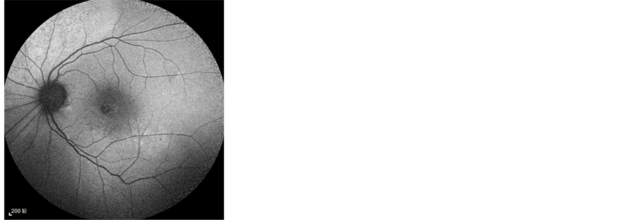 (b)
(b) 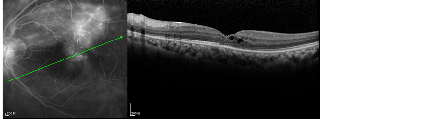 (c)
(c)
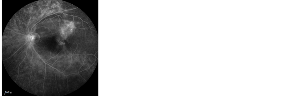
Figure 1. A case of retinal vasculitis
图1. 视网膜血管炎一例
(a) 视网膜血管炎FAF显示黄斑中心凹颞侧片状高荧光改变;(b) FFA晚期显示黄斑区及其上方片状高荧光积存;(c) FFA联合OCT检查显示黄斑区高荧光积存区可见网膜层间囊样改变
 (a)
(a)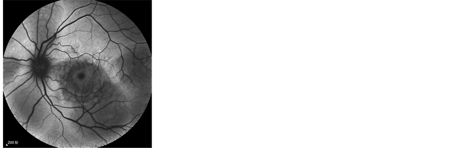 (b)
(b)
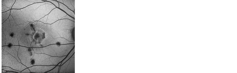
Figure 2. A case of Vogt-Koyanagi Harada disease
图2. Vogt-小柳原田病一例
(a) 原田病(急性期)患者行FAF检查显示后极部多囊样视网膜脱离区呈低自发荧光(因为水肿区视网膜下液体遮蔽来自RPE的自发荧光),而脱离区出现的高自发荧光提示RPE代谢障碍; (b) 激素治疗1个月后,FAF显示囊样低荧光明显减轻,异常高荧光区减少
4. 讨论
随着年龄的增长,RPE中因脂褐质和自发荧光基团积聚而产生自发荧光(AF)。脂褐质及其主要荧光
 (a)
(a)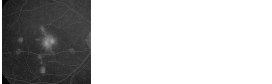 (b)
(b)  (c)
(c)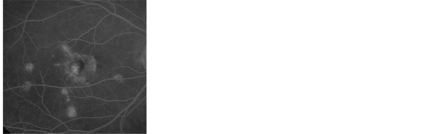 (d)
(d)
Figure 3. A case of multifocal choroiditis with CNV
图3. 多灶性脉络膜炎伴发CNV一例
(a) 多灶性脉络膜炎伴黄斑区CNV患者FFA示后极部多处小灶性透见高荧光,黄斑区CNV荧光明显渗漏;(b) FAF显示后极部多处小灶性低荧光(与FFA透见荧光一致),黄斑区出血呈遮蔽荧光,其周围病灶边缘可见高荧光;(c) 3月后复诊FFA显示黄斑区CNV得到控制;(d) 复诊FAF显示黄斑区弱高荧光降低,萎缩灶(低荧光)扩大,提示病情趋于稳定
Table 2. The contrastive evaluation of the image feature in FAF between before treatment and after treatment for posterior uveitis
表2. 对比评价后葡萄膜炎治疗前后眼底自发荧光的表现
团N-视黄基-乙醇胺(A2-E)对RPE细胞具有包括溶酶体降解等多重破坏作用。因此,RPE-AF增加预示着地图状萎缩区内将要发生细胞死亡,RPE-AF减低主要发生在RPE细胞减少或萎缩的区域。一般而言,AF的差异代表着RPE细胞中的代谢变化,因此累及RPE病变的疾病常伴有异常AF表征[9] 。后葡萄膜炎是累及脉络膜、视网膜及玻璃体的炎症性疾病,多表现有RPE病变,且其常伴发有黄斑水肿,由于水肿主要位于外丛状层及内核层,水肿的囊腔推挤色素使黄斑色素密度降低,故囊腔所在区域在FAF中因缺乏吸收激发光的物质而表现为高荧光[10] 。本研究中,我们对后葡萄膜炎黄斑水肿[11] [12] 及后葡萄膜炎治疗前后疗效评价等进行了分析。结果显示:1) 在观察后葡萄膜炎黄斑水肿中,FFA/ICGA显示黄斑区呈高荧光积存(即提示黄斑水肿)的患眼在FAF中均可见黄斑区强荧光,另外存在2眼(多灶性脉络膜炎)FAF显示黄斑区强荧光而FFA/ICGA中并未呈现强荧光,考虑可能为病变进展期脂褐质聚集、迁徙的含有脂褐素的RPE细胞或巨噬细胞所致,而在眼底血管造影中并无高荧光表现;2) 在观察后葡萄膜炎治疗前后的变化中,FAF显示治疗1月后异常高荧光较前减弱,考虑是由于RPE的损害得到控制或进行性萎缩,RPE的代谢产物脂褐质堆积量逐渐减少或RPE逐步凋亡,使能够吸收产生高荧光的物质明显减少,则AF信号降低,呈较低荧光改变,同时黄斑水肿也有效减轻,黄斑色素密度增加也使AF信号降低;另外,由于网膜下液遮蔽脂褐质的自发荧光而使之呈低荧光表现,经治疗积液吸收后低荧光也随之减退。由于FAF用于临床时间较短,患者接受度有限,限制了研究中样本的数量,研究结果尚不够全面,我们将在今后的临床实践中继续收集样本扩大研究。
综上所述,本研究资料显示:FAF在评估后葡萄膜炎黄斑水肿及评价后葡萄膜炎治疗前后疗效分析等方面具有无创、快速、准确、可重复等优点,有特殊的临床应用价值。这提示在临床工作中,对于不能或不能及时行眼底血管造影等检查的患者,FAF以其安全无创、快速有效的特点可用于后葡萄膜炎等疾病的辅助诊断、随访观察和治疗评估。
基金项目
江苏省“青蓝工程”项目,项目编号:苏教师(2012)39号。

NOTES
*通讯作者。