Abstract:
In order to obtain the high-quality images, this work improved the traditional full-field optical coherence tomography (full-field OCT) which was based on the Michelson interferometer and visible light. Unlike the common light used in the most OCT, this paper design to substitute the halogen lamp with the infrared light. This design could improve the resolution and the detecting depth, and could achieve the noninvasive optical imaging. This paper provided a practical way for the noninvasive live-cell imaging based on the full-field OCT.
1. 引言
光学相干层析(optical coherence tomography, OCT)成像技术的核心实际上是一个迈克尔逊干涉仪 [1] ,其利用宽带光源的低相干性,通过测量从生物组织或材料内部不同深度的后向散射光和参考光的干涉光强来得到样品内部微观结构的层析图像。OCT是基于光学方法制作出的生物传感器,具有非接触、无损伤,高分辨率,成本低,快速成像和实时监测等优点。OCT技术有着广泛的应用,目前已应用于眼科,皮肤科,心血管,消化道,呼吸系统,口腔和牙齿组织,以及活体细胞的观察 [2] - [8] 。
全场光学相干层析成像(FF-OCT)系统是OCT技术的一个分支。对于全场OCT系统而言,光源的选择非常重要,光源的中心波长越长,OCT系统的纵向分辨率就越高,成像深度也越深。我们设计的红外光源的中心波长为1000 nm,高于目前广泛运用的各种光源,可以提高系统的纵向分辨率和成像深度。另外,一般光源照射生物样品,尤其是波长500纳米以下的蓝紫光,对生物样品有很大的杀伤性,能够损伤DNA结构。而在600~1300纳米之间的近红外光谱范围内,生物组织具备良好的透光性,对光的吸收小,且近红外技术能够实现真正意义上的无损检测。所以,本文设计的近红外光源,理论上可以实现对生物组织的无损成像,这就消除了在活体探测过程中对生物样品的光损伤,适于对活体组织或细胞长时间的观察。
2. 全场OCT装置
如图1所示,在两条臂中加入了两个相同的显微物镜。这种干涉仪又叫做Linnik干涉显微系统。通过本文设计的红外光源发出的光穿过透镜1后经分束镜被分为两束,分别射向参考臂和样品臂,随后投射在两个相同型号的显微物镜的后焦面上。从两臂反射回的光发生干涉,干涉信号由探测器接收。参考臂由压电陶瓷(piezoelectric ceramics, PZT)调制,通过控制PZT的电压来控制位移量的变化,使参考臂进行移动。需要观察的生物组织样品则在活细胞培养系统中放置。样品臂由步进电机做纵向扫描,则可获得样品三维信息。
3. 系统组成
3.1. 光源
本文选用卤钨白炽灯作为热辐射红外光源。首先卤钨白炽灯作为热光源,具有低时间相干性和低空间相干性,这样既保证了高分辨率,又在很大程度上抑制并行探测中出现的空间串扰(cross-talk),从而降低了图像的散斑噪声。而且相比于其他OCT系统中所使用的光源,如超辐射发光二极管(the superluminesent diode, SLD)、超快激光器等,其价格十分便宜,能够降低OCT系统的成本。本文设计的红外光源
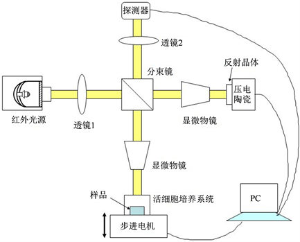
Figure 1. Schematic diagram of full-field OCT
图1. 全场OCT装置示意图
由卤钨白炽灯、聚光反射镜、滤光片组成。发光面处于聚光镜的焦点上,光束成平行光。如图2所示。
OCT图像的纵向分辨率主要与光源的中心波长和带宽有关。
其中,
是光源的中心波长,
是光源的带宽。
图3为卤素灯与本文设计的红外光源光谱对比图。红外光源其中心波长为1000 nm,带宽为150 nm。
理论纵向分辨率为:
。而如全场OCT系统中一般用到的中心波长为600~700
nm,带宽150~300 nm的光源,纵向分辨率为0.7~1 μm [9] 。由此可见,本文设计的光源大大提高了纵向分辨率。
3.2. 透镜
一般的OCT系统使用的都是可见光,所以该系统的透镜也都是针对可见光的,这明显不符合本文红外光源的需求。所以本文设计使用了菲涅尔透镜作为传统透镜的替代品。菲涅尔透镜简单的说就是在透镜的一侧有等距的齿纹,通过这些齿纹可以达到对制定光谱范围的光带通(反射或折射)的作用。与传统透镜相比,菲涅尔透镜具有重量轻,材料来源丰富、成本低、制作方便、厚度薄等特点 [10] 。
3.3. 分束镜
中心波段为可见光波段的分束镜亦不能满足本套系统的要求,我们设计使用红外分束镜来将从光源处接收到的红外光分为两束,分别进入参考臂和样品臂。
3.4. 探测器
由于在红外波段对生物样品进行成像,二维探测器也需从以Si为基底的CCD转换为探测红外波段的以InGaAs为基底的CCD,与此同时,系统的成像深度也得到了提高 [11] 。
本文设计的全场OCT系统相关光学参数见表1。
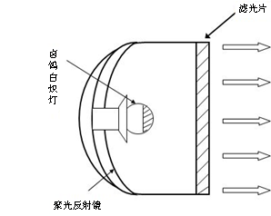
Figure 2. Structure chart of infrared light composed of tungsten halogen lamps
图2. 由卤钨白炽灯组成的红外光源结构图
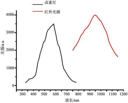
Figure 3. Spectra of Halogen lamp and infrared light designed in this paper
图3. 卤素灯与本文设计的红外光源光谱对比图

Table 1. Optical parameters of our designed full-field OCT
表1. 本文设计的全场OCT系统相关光学参数
4. 结论
本文主要针对全场OCT系统装置的光源进行了改良研究,设计了以卤钨白炽灯作为红外光源的全场OCT成像系统。理论上提高了该系统成像的分辨率和探测深度,并且做到了对生物样品的无损成像,为主要应用于生物组织活体成像的全场OCT系统的改进提供了切实可行的方法。
基金项目
辽宁省自然科学基金(2015020715),沈阳师范大学优秀人才支持计划(BS201433),沈阳师范大学自主科研计划(L201514)和沈阳师范大学博士启动计划(51600209)资助。