1. 引言
食管纤维血管性息肉(Esophageal fibrovascular polyp, EFVP)是一种少见的良性肿瘤性病变,1990年世界卫组织(WTO)国际肿瘤组织学分型将纤维瘤、纤维上皮息肉、带蒂脂肪瘤、纤维脂肪瘤、粘液纤维瘤等统称为纤维血管性息肉 [1] 。目前病因不明,早期症状不明显,病变长大后,患者临床症状较痛苦,且有呼吸困难、窒息等严重症状,并有恶变可能。巨大纤维血管性息肉较少见,文献报道较少,为提高对本病的认识、诊断,为临床手术提供详细影像学支持,现结合文献复习报告如下。
2. 临床资料
患者男,45岁,主诉间断恶心、呕吐10年余,加重1月。患者于6年前发现呕吐时可有长蒂息肉样肿物由咽部呕出,呕吐后可回纳消失。外院检查:喉镜示反流性咽喉炎;胃镜示食管距门齿20~30 cm处左侧壁见粘膜下肿物,游动,性质待定;增强CT示多发食道粘膜下巨大囊肿?粘膜脱垂?于2016年1月25日入我院消化内科门诊就诊,门诊以“食管占位性质待定”收住消化内科,入院后上消化道钡餐检查示(图1):食管上段、中段局限性充盈缺损,结合临床病史,考虑食道息肉。胃镜示(图2):食管巨大息肉样肿物。胸部CT平扫加增强并多平面重建示(图3):食道于胸1椎体水平至胸9/10椎间隙水平见长条形混杂密度影,长度约18.0 cm,边界清晰,边缘光整,以长蒂与食道上段相连,蒂基底部约宽1.5 cm,长约2.3 cm,另病变中段(平胸4椎体水平)见软组织密度影与其相连,大小约1.3 cm × 3.9 cm,连接部宽约0.9 cm,增强后可见强化,考虑巨大息肉样病变。
患者2016年2月3日在我院消化内科内镜中心行食管巨大息肉样肿物内镜下切除术,术中距门齿15 cm至距门齿37 cm可见一条形巨大病变,蒂位于近食管入口处,使用导丝将病变根部扎紧,钛夹于导丝旁夹紧瘤体,电刀切除,切除物使用圈套器套紧自口中取出,甲醛固定,送病理。术后病理示(图4~6):灰白色长条状肿物一个,大小18.0 cm × 2.7 cm × 2.0 cm (上下径 × 左右径 × 前后径),表面灰白色,切面呈灰黄色胶冻样,所连灰褐色息肉样物一块,大小1.3 cm × 6.1 cm。病理诊断:(食管)良性纤维血管性息肉。患者术后恢复良好,病情稳定,术后第3天出院。
3. 讨论
3.1. 发病原因
食管纤维血管性息肉是一种少见的良性肿瘤性病变,可发生于任何年龄,男性多于女性,目前报道最小的患者为5个月 [2] 。食管纤维血管性息肉早期症状不明显,这也是导致巨大纤维血管性息肉形成的原因,病变由纤维组织、脂肪组织及血管等构成,表面为正常分化的鳞状上皮覆盖,食管纤维血管性

Figure 1. Gastrointestinal imaging, Esophageal upper section can be seen filling defect, Smooth surface, Visible separate
图1. 消化道造影检查,食管中上段可见充盈缺损,病变表面光滑,可见分叶
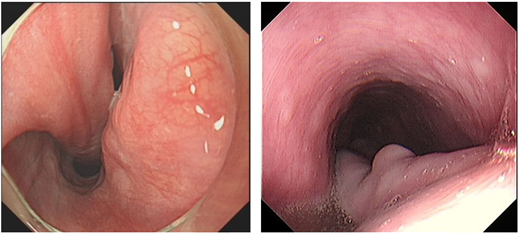
Figure 2. Endoscopic examination of the esophagus and esophageal wall can be seen within the polypoid mass
图2. 胃镜检查食管内可见以蒂与食管壁相连的息肉样肿物

Figure 3. Esophagus see the length of about 18 cm long strip of mixed mass, clear boundaries, neat edge, with a long strip connected with the upper esophagus, the middle of the lesion (Flat chest 4 vertebral level) See soft tissue mass connected to it, Size about 1.3 × 3.9 cm, The width of the connecting portion is about 0.9 cm, CT scan inhomogeneous enhancement
图3. 食道内见长度约18 cm长条形混杂密度影,边界清晰,边缘光整,以长蒂与食道上段相连,病变中段(平胸4椎体水平)见软组织密度影与其相连,大小约1.3 × 3.9 cm,连接部宽度约0.9 cm,CT增强扫描食管内肿物不均匀强化

Figure 4. The lesions are rich in capillaries, Surface squamous epithelium, HE 40×
图4. 病变内富含毛细血管,表面为鳞状上皮HE 40×

Figure 5. Squamous epithelium and capillaries HE 1000×
图5. 鳞状上皮与毛细血管HE 1000×

Figure 6. Lesion size is 18.0 cm × 2.7 cm × 2.0 cm, the size of the attached tumor was 1.3 cm × 6.1 cm
图6. 病灶切除大体大小为18.0 cm × 2.7 cm × 2.0 cm,分叶大小为1.3 cm × 6.1 cm
息肉通常起源于颈段食管邻近环咽肌的黏膜下层 [3] ,在生长过程中受到食管蠕动挤压而形成较大的带蒂腊肠样肿物。食管纤维血管性息肉可发生癌变,但发生率极低,其中脂肪成分可恶变成脂肪肉瘤,有报道此恶变成分还可伴有横纹肌分化,鳞状上皮可恶变成鳞状细胞癌,小的息肉还可能发展成腺癌 [4] 。
食管纤维血管性息肉诊断可依据X线钡剂检查、消化内窥镜和CT等检查方式,X线钡剂检查可发现病变形态及位置;内镜可直接观察息肉大小、位置、形态,同时还可钳取组织病检或进行镜下手术操作;CT可检测出肿物的主要组成成分、位置、与食管关系,有时在增强CT上可见到息肉内部由根蒂部发出的营养血管。
本例患者临床症状为恶性、呕吐,并可吐出息肉样肿物(图7),在消化道造影检查中显示病灶巨大,占位效应明显,在CT增强并三维重建下进一步确定病变大小、形态、血供情况及与食管关系。术前影像学检查为最终内镜切除病灶提供了详细、准确的影像学基础,术前准备充分,术后病变大体形态与术前影像学检查所示病灶形态高度一致,最终术前影像学诊断与最后病理结果一致。
3.2. 鉴别诊断
本病通常可依据患者临床症状,结合食道造影、消化道内窥镜及胸部CT等影像学检查明确诊断。本病需与食管恶性病变及贲门失弛缓症相鉴别。
3.3. 治疗
本患者在进行了详细的影像学检查后,通过消化内镜进行手术切除。
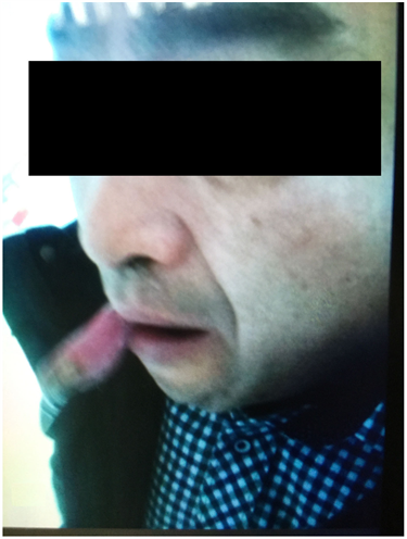
Figure 7. The polyp is spit out by the mouth of the patient
图7. 息肉由患者口中吐出
4. 结论
因病变通常体积较大易引起不同程度的吞咽困难和食物梗塞,甚至因息肉的反流吸入气管,而引起呼吸困难、窒息等严重并发症。故一经发现,应当及时手术治疗。通过详细的影像学检查后,确定肿物构成成分及与食管的位置关系,若主要由纤维、脂肪组织构成,其间仅含少量血管,出血的可能性减小,且以蒂与食管相连,便可考虑内镜下切除 [5] ,若肿物基底宽阔,因根据其所在部位,选择经颈部或胸切除。因此术前通过影像学检查明确根蒂部位置、宽度及血供情况极为重要。因食管巨大纤维血管性息肉较为罕见,我院病例收集仅此一例,故此研究尚存在一定局限性。
NOTES
*通讯作者。