胸腔镜下肺切除术伴部分型肺静脉异位连接的个案报道
A Case Report of Thoracoscopic Pneumonectomy with Partial Anomalous Pulmonary Vein Connection
DOI: 10.12677/HJS.2021.104012, PDF, HTML, XML, 下载: 406  浏览: 1,005 
作者: 衣腾飞*, 邵盛腾, 李 烁, 刘玉洪#:青岛大学附属医院胸外科,山东 青岛;秦 坤:青岛大学附属医院急诊科,山东 青岛
关键词: 肺癌部分型肺静脉异位连接手术治疗Lung Cancer Partial Anomalous Pulmonary Venous Connection Operative Treatment
摘要: 部分型肺静脉异位连接(Partial anomalous pulmonary venous connection, PAPVC)是指4支肺静脉中的1条或数条(非全部)肺静脉未与左心房直接连接,而与右心房或体静脉相连的一种先天性心血管畸形。属于比较少见的先天性心血管异常性疾病,而肺癌合并PAPVC的案例更是罕见。现报道青岛大学附属医院收治的1例左肺上叶腺癌合并同侧孤立性PAPVC的病例。
Abstract: Partial anomalous pulmonary venous connection (PAPVC) is a congenital cardiovascular malformation in which one or more (but not all) of the 4 pulmonary veins are not directly connected to the left atrium, but connected to the right atrium or body vein. It is a relatively rare congenital abnormal cardiovascular disease, and the case of lung cancer complicated with PAPVC is even rarer. A case of left upper lobe adenocarcinoma complicated with ipsilateral isolated PAPVC is reported in the Affiliated Hospital of Qingdao University.
文章引用:衣腾飞, 邵盛腾, 秦坤, 李烁, 刘玉洪. 胸腔镜下肺切除术伴部分型肺静脉异位连接的个案报道[J]. 外科, 2021, 10(4): 69-74. https://doi.org/10.12677/HJS.2021.104012

1. 前言

部分型肺静脉异位连接(Partial anomalous pulmonary venous connection, PAPVC)是指4支肺静脉中的1条或数条(非全部)肺静脉未与左心房直接连接,而与右心房或体静脉相连的一种先天性心血管畸形。其临床症状的严重程度取决于肺静脉异位引流所致的左向右分流程度:分流量小的患者可无症状;而畸形引流静脉粗大则常出现胸痛、心悸、呼吸困难等症状,部分可伴有肺动脉高压。对于少量左向右分流的无症状患者无须手术治疗,但患者进行肺切除术时则需同时处理畸形静脉,否则会加重左向右分流甚至出现右心衰竭 [1]。本文介绍的是左肺上叶腺癌合并同侧孤立性PAPVC的病例。

2. 病例报告

患者为63岁老年男性,因“查体发现左肺上叶结节2年余”入院。自诉无胸闷、憋气,无咳嗽、咳痰、咯血,无胸痛,无饮水呛咳及声音嘶哑,无心悸、乏力、头晕等症状。入院后复查胸部CT显示:“左肺上叶尖段见不规则混合磨玻璃斑片影,大小约20 * 15 * 10 mm,可见血管穿行,边缘分叶。纵隔淋巴结大小在正常范围”(见图1)。超声心动图检查未见房间隔或室间隔缺损,心血管活动正常,PASP为26 mmHg;肺功能大致正常,FEV1/FVC%预计值为76.05%;血气分析显示PaO2为88 mmHg,PaCO2为39 mmHg,氧饱和度为96%。查体颈静脉无怒张,心率86次/分,律齐,各瓣膜听诊区未闻及病理性杂音。双下肢无水肿。既往高血压病史10年余,平素规律服用降压药物,血压控制在120/80 mmHg;吸烟史45年余,无饮酒史;否认家族中有遗传倾向性及传染性疾病。该病例报道已获得患者及家属的知情同意。

经评估,考虑结节恶性可能性大,加上舌段因解剖学特点难以保留,决定行胸腔镜下左肺上叶切除+系统性淋巴结清扫术。术中探查未见胸水和粘连,开始游离前肺门,发现左肺上叶肺静脉并未汇入心包,而是在膈神经后方骑跨主动脉弓汇入左头臂干静脉(见图2图3),下叶肺静脉连接正常。再次查看患者胸部CT,发现左侧主动脉弓旁出现一个异常血管影,最终汇入左头臂干静脉(见图4图5)。然后依次游离并切割A1 + 2c支、A1 + 2a + b支和A3支;充分游离左肺上叶变异静脉,使用ENDO-GIA45#棕钉闭合切割靠近头臂静脉汇合处的上肺静脉,以避免残端过长(如图6);最后游离并切断上叶支气管。术后病理结果示:(左肺上叶)腺泡型浸润性腺癌(周围型),部分区域呈乳头状生长,约占10%。

Figure 1. Chest computed tomography shows mixed ground-glass nodule located at the S1 segment of the left lung

图1. 胸部CT显示混合磨玻璃结节位于左肺上叶尖段
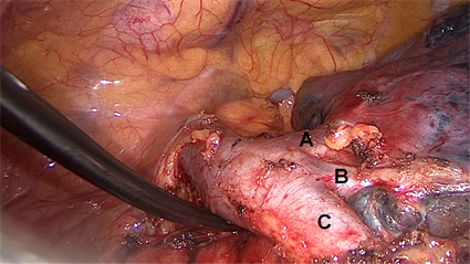
Figure 2. A, B and C are generic branches of the pulmonary vein in the upper lobe of the left lung. A + B: V1+2a-d + V3a + b + c. C: V4+5

图2. A,B and C是左肺上叶肺静脉的属支。A + B: V1+2a-d + V3a + b + c. C: V4+5
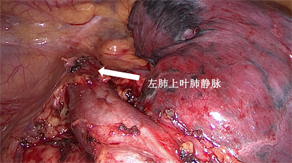
Figure 3. The pulmonary vein of the left upper lobe flowed into the left brachiocephalic trunk vein (arrow)

图3. 左肺上叶肺静脉汇入左头臂干静脉(箭头)
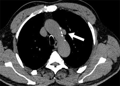
Figure 4. The mediastinal window of chest computed tomography demonstrates an abnormal vascular shadow near the left aortic arch (arrow), and the abnormal vascular shadow is the left superior pulmonary vein (LSPV)

图4. 胸部CT纵隔窗显示主动脉弓旁见一异常血管影(箭头),此异常血管即为左肺上叶肺静脉
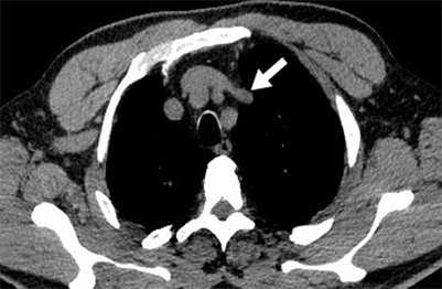
Figure 5. The left superior pulmonary vein (LSPV) eventually drains into the left brachiocephalic vein (BCV) (arrow)

图5. 左肺上叶肺静脉最终汇入左头臂干静脉(箭头)
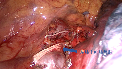
Figure 6. The left superior pulmonary vein (LSPV) was cut and closed (arrow)

图6. 闭合切断左肺上叶肺静脉(箭头)

3. 讨论

PAPVC是一种比较少见的先天性心血管异常性疾病。胚胎发育过程中,肺静脉丛未与肺静脉原基连接而与内脏静脉(如右前、左前主要静脉,脐卵黄静脉等)相连,导致一部分肺静脉开口于右心房或通过腔静脉系统再注入右心房 [2] [3]。仅存在于0.5%~0.7%的人群中 [2] [4],而肺癌合并无房间隔缺损的左侧PAPVC则更为罕见。

Black [5] 等人于1992年首次报道了一例肺癌合并PAPVC的病例,肿块位于右肺门而PAPVC位于左肺上叶,实施右全肺切除术后不久,由于左向右分流量增加患者出现右心衰竭,随即迅速采取心肺旁路术也未能挽救患者生命。这也是唯一一例公开报道患者死于肺切除术后的肺动脉高压。因此,肺癌合并PAPVC的患者中,两者位于同一肺叶时,肺切除术可同时解决肺癌和静脉异位引流畸形;但当两者不在同一肺叶时,行全肺或双肺叶切除会增加左向右分流量,进而导致心力衰竭 [5],这类患者在行肺切除术的同时纠正变异静脉是必要的。

幸运的是,本病例是肺癌和PAPVC均位于左肺上叶且不伴任何心内畸形,即孤立性部分型肺静脉异位引流(isolated partial pulmonary venous drainage)。上叶肺静脉流入头臂静脉,氧合的动脉血直接进入体循环静脉,致使静脉血变为混合动脉血,再经右心房及右心室流入肺动脉,形成无效循环,上叶的肺换气变成了无效做功;但患者无任何症状且无肺动脉高压。因此,肺叶切除术是完全安全可行,并且一举处理了变异静脉。

PAPVC类型较多,右侧肺静脉常异位引流至上腔静脉、下腔静脉、右心房、奇静脉、门静脉及肝静脉 [5] [6] [7] [8];左侧肺静脉常异位引流至左无名静脉、冠状静脉窦及半奇静脉 [9] - [14]。2002年,Takei [11] 报道了一例左肺下叶肺腺癌合并上叶肺静脉异位连接且伴肺动脉高压的病例。采取解剖性左肺下叶切除 + 淋巴结清扫术,同时,将异常的左肺上叶肺静脉与左头臂静脉充分游离并切断上肺静脉,然后将上肺静脉与近端下肺静脉进行端端吻合;术后无任何心血管并发症发生。

总的来说,肺癌合并PAPVC的案例十分罕见,且大多在术中被发现。此患者较幸运,无肺动脉高压且肺切除术可完全解决肺癌和变异静脉;但肺癌和PAPVC不在同一肺叶时,手术较为复杂且存在心血管并发症风险。因此,胸外科医生在术前要有意识地考虑到PAPVC,这样才会在患者的影像学检查上发现变异血管,以制定安全合理的手术方案。

NOTES

*第一作者。

#通讯作者。

参考文献

[1] Inafuku, K., et al. (2016) Thoracoscopic Lobectomy for Lung Cancer in a Patient with a Partial Anomalous Pulmonary Venous Connection: A Case Report. Journal of Cardiothoracic Surgery, 11, 113.
https://doi.org/10.1186/s13019-016-0527-7
[2] Dillman, J.R., Yarram, S.G. and Hernandez, R.J. (2009) Imaging of Pulmonary Venous Developmental Anomalies. AJR. American Journal of Roentgenology, 192, 1272-1285.
https://doi.org/10.2214/AJR.08.1526
[3] Brody, H. (1942) Drainage of the Pulmonary Veins into the Right Side of the Heart. America Heart Journal, 33, 221.
[4] Zylak, C.J., Eyler, W.R., Spizarny, D.L. and Stone, C.H. (2002) Developmental Lung Anomalies in the Adult: Radiologic-Pathologic Correlation. Radiographics, 22, S25-S43.
https://doi.org/10.1148/radiographics.22.suppl_1.g02oc26s25
[5] Black, M.D., Shamji, F.M., Goldstein, W. and Sachs, H.J. (1992) Pulmonary Resection and Contralateral Anomalous Venous Drainage: A Lethal Combination. The Annals of Thoracic Surgery, 53, 689-691.
https://doi.org/10.1016/0003-4975(92)90336-3
[6] Sasaki, H., et al. (2006) Right Partial Anomalous Pulmonary Venous Connection Found during Lobectomy for Coexisting Lung Cancer and Tuberculosis: Report of a Case. Clinical Lung Cancer, 7, 350-352.
https://doi.org/10.3816/CLC.2006.n.018
[7] Heineman, D.J., van den Aardweg, J.G. and Schreurs, W.H. (2015) A Rare Case of Anomalous Venous Drainage of the Right Upper Lobe. Journal of Thoracic Disease, 7, E502-E504.
[8] Mikubo, M., et al. (2013) Pulmonary Resection of Lung Cancer in a Patient with Partial Anomalous Pulmonary Venous Connection. The Annals of Thoracic Surgery, 95, 1799-1801.
https://doi.org/10.1016/j.athoracsur.2012.10.033
[9] Sakurai, H., et al. (2005) Left Pneumonectomy for Lung Cancer after Correction of Contralateral Partial Anomalous Pulmonary Venous Return. The Annals of Thoracic Surgery, 79, 1778-1780.
https://doi.org/10.1016/j.athoracsur.2003.10.092
[10] Takei, H., Suzuki, K., Asamura, H., Kondo, H. and Tsuchiya, R. (2002) Successful Pulmonary Resection of Lung Cancer in a Patient with Partial Anomalous Pulmonary Venous Connection: Report of a Case. Surgery Today, 32, 899-901.
https://doi.org/10.1007/s005950200176
[11] Asakura, K., et al. (2014) Partial Anomalous Pulmonary Venous Connection Associated with Lung Cancer in the Same Lobe: Report of a Case. Annals of Thoracic and Cardiovascular Surgery, 20, 457-460.
https://doi.org/10.5761/atcs.cr.12.01975
[12] Shapiro, M., Bittermann, C. and Flores, R.M. (2014) Anomalous Pulmonary Vein Drainage Is Not a Contraindication for VATS Lobectomy. The Thoracic and Cardiovascular Surgeon, 62, 255-257.
https://doi.org/10.1055/s-0032-1328926
[13] Miwa, K., Takamori, S., Hayashi, A., Fukunaga, M., Ikeda, K. and Shirouzu, K. (2004) Incidental Partial Anomalous Pulmonary Venous Connection in Left Lung Cancer. The Japanese Journal of Thoracic and Cardiovascular Surgery, 52, 189-190.
https://doi.org/10.1007/s11748-004-0105-0
[14] Kawagoe, I., et al. (2017) A Combination of a Partial Anomalous Pulmonary Venous Connection (PAPVC) and a Lung Tumor Requiring Pulmonary Resection. Journal of Cardiothoracic and Vascular Anesthesia, 31, 274-278.
https://doi.org/10.1053/j.jvca.2016.06.011